Introduction
The human heart is a tireless engine, beating over 100,000 times a day to sustain life. Yet, cardiovascular disease remains one of the most significant health challenges of our time. Often, heart conditions develop quietly, hidden beneath the surface until a sudden event forces us to confront our fragility. This is why the evolution of cardiac diagnostics—specifically the ability to peer inside the living, beating heart—has become a cornerstone of modern medicine.
When searching for “cardiac imaging centers near you,” you are looking for more than just a place to get a test. You are seeking a facility that blends cutting-edge technology with the human expertise required to interpret complex data accurately. Navigating these options can feel overwhelming, which is why resources like BESTCARDIACHOSPITALS serve as a vital compass for patients and families alike.
What is Cardiac Imaging?
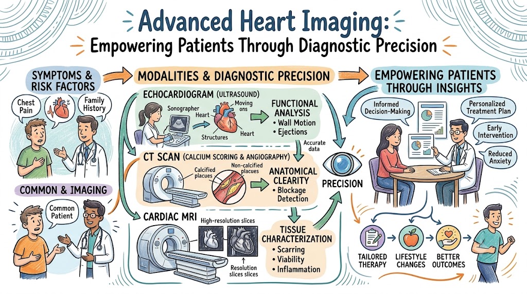
Cardiac imaging refers to the use of non-invasive or minimally invasive technology to visualize the heart and its associated blood vessels. Unlike standard diagnostic tests that look at the heart’s electrical rhythm (like an ECG) or broad physical health, cardiac imaging provides a visual blueprint of the heart’s anatomy and function.The purpose of these exams is to answer specific questions: Is the heart muscle pumping effectively? Are the valves opening and closing as they should? Is there plaque buildup in the coronary arteries? By providing high-definition imagery, these tests allow cardiologists to view the heart in real-time, helping them understand not just how the heart is functioning, but exactly why a patient might be experiencing symptoms like chest pain, dizziness, or fatigue.This field has moved far beyond simple X-rays. Today, we utilize sophisticated magnetic resonance, computerized tomography, and ultrasonic waves to build a complete diagnostic picture. For the patient, this means a safer, faster, and more accurate diagnostic process that is central to the practice of preventive cardiology.
Why Cardiac Imaging is Important
The primary value of cardiac imaging lies in its ability to facilitate early disease detection. Many heart conditions, such as cardiomyopathy or early-stage coronary artery disease, do not present with symptoms until they reach an advanced state. Imaging allows clinicians to intercept these conditions.
Furthermore, accurate diagnosis is the foundation of effective treatment. Without imaging, a doctor might treat symptoms; with imaging, a doctor treats the root cause. For example, knowing exactly which valve is malfunctioning allows a surgeon to plan a procedure with precision, minimizing risks and recovery time. Additionally, imaging plays a critical role in monitoring the success of an ongoing treatment plan, ensuring that the heart is responding positively to medication or lifestyle changes.
Table 1: Benefits of Cardiac Imaging
| Benefit | Why It Matters |
| Early Detection | Identifies disease sooner, improving prognosis |
| Accurate Diagnosis | Ensures targeted, effective treatment plans |
| Risk Assessment | Allows for preventive care and lifestyle modifications |
| Treatment Monitoring | Tracks progress and adjusts interventions |
| Personalized Care | Matches treatment to the patient’s specific anatomy |
Types of Cardiac Imaging Tests
Advanced imaging centers provide a suite of tools, each designed to capture a unique aspect of heart health.
Echocardiography
This is essentially an ultrasound of the heart. It uses sound waves to create live images of your heart in motion. It is the gold standard for evaluating heart valve function, wall thickness, and overall pumping efficiency.
Cardiac CT Scan
A Cardiac Computed Tomography (CT) scan uses X-rays to create detailed 3D images of your heart and blood vessels. It is especially useful for “coronary calcium scoring,” which measures the amount of calcified plaque in your arteries, providing a window into your long-term heart attack risk.
Cardiac MRI
Using magnetic fields rather than radiation, a Cardiac Magnetic Resonance Imaging (MRI) scan provides the most detailed look at heart muscle tissue. It is essential for diagnosing inflammatory heart conditions, assessing scarring after a heart attack, and evaluating complex congenital heart defects.
Nuclear Cardiology Imaging
This involves the use of a small amount of radioactive tracer to visualize blood flow to the heart muscle. It is highly effective at determining whether a specific part of the heart is receiving enough blood during rest and exercise.
Stress Testing
While often functional in nature, modern stress testing often incorporates imaging (like nuclear or echo) to observe how the heart behaves under physical or chemical stress, helping to identify blockages that only appear when the heart is working hard.
Conditions Detected Through Cardiac Imaging
Cardiac imaging is the “eyes” of the cardiologist. It is instrumental in detecting:
- Coronary Artery Disease: Identifying blockages or plaque in the vessels that supply the heart.
- Heart Failure: Assessing if the heart muscle is weakened or unable to pump efficiently.
- Cardiomyopathy: Evaluating structural diseases of the heart muscle.
- Valve Disorders: Determining if valves are leaking (regurgitation) or too narrow (stenosis).
- Congenital Heart Disease: Visualizing structural abnormalities present from birth.
- Pericardial Disease: Looking at the protective sac surrounding the heart for inflammation or fluid buildup.
Who Should Consider Cardiac Imaging?
Cardiac imaging is not reserved only for those with a known heart condition. It is a vital tool for risk stratification. You should consider cardiac imaging if you fall into any of the following categories:
- Symptomatic Patients: Individuals experiencing unexplained chest pain, shortness of breath, palpitations, or fainting spells.
- Family History: Those with a strong family history of premature heart disease, as genetic predispositions may require early, non-invasive surveillance.
- Chronic Conditions: Individuals with diabetes, hypertension, or high cholesterol, as these conditions are “silent” drivers of coronary artery disease.
- Athletes: High-performance athletes may undergo screening to rule out structural issues that could predispose them to sudden cardiac events.
- Older Adults: As part of a preventive health strategy to assess long-term heart health.
How to Prepare for Cardiac Imaging Tests
Preparation varies depending on the specific test, but most cardiac imaging is designed to be as non-invasive as possible.
- Medical History: Be prepared to provide a detailed list of your medications, allergies, and any history of implants (especially for MRI).
- Fasting: For tests like a Cardiac CT or Nuclear Imaging, you may be asked to fast for several hours beforehand to ensure clear imagery.
- Medication Guidance: Some tests require you to pause or adjust heart medications (such as beta-blockers) prior to the appointment. Always clarify this with your clinic.
- Expectations: Most tests are painless. You will be asked to lie still, and you may hear clicking sounds (in MRI) or be asked to hold your breath for short intervals (in CT).
How to Choose the Best Cardiac Imaging Center
When you look for cardiac imaging centers near you, prioritize facilities that offer a comprehensive diagnostic environment.
- Board-Certified Specialists: Ensure the imaging is interpreted by radiologists or cardiologists with sub-specialty training in cardiovascular imaging.
- Technological Infrastructure: High-quality imaging requires modern hardware. Look for centers that utilize the latest MRI and CT technology.
- Accreditation: Check if the center is accredited by national bodies, which ensures they follow strict safety and quality protocols.
- Comprehensive Care: The best hospitals often combine imaging with emergency and interventional services. This ensures that if a scan reveals a critical finding, you are already within a system that can provide immediate, life-saving care.
- Patient Support: Look for centers that provide clear explanations, offer support throughout the imaging process, and maintain efficient systems for sharing results with your primary cardiologist.
Risks, Limitations & Safety of Cardiac Imaging
While cardiac imaging is generally safe, it is important to be an informed patient.
- Radiation: Tests like Cardiac CT scans involve small amounts of ionizing radiation. Modern machines use low-dose protocols to minimize this exposure.
- Contrast Dye: Some exams use a contrast agent to highlight blood vessels. Patients with poor kidney function or specific allergies must discuss this with their doctor beforehand.
- MRI Safety: Cardiac MRI uses powerful magnets. If you have metal implants, pacemakers, or certain medical devices, this must be disclosed immediately, as it may be unsafe or require a special protocol.
- Expert Interpretation: The technology is only as good as the person reading the images. This is why choosing a center with board-certified imaging specialists is critical to avoid false positives or negatives.
Recovery & Follow-Up After Imaging
One of the greatest benefits of modern cardiac imaging is the speed of recovery.
- Post-Test: For most imaging, you can resume normal activities immediately. If sedation was used (rare, but possible for some MRI protocols), you may need someone to drive you home.
- Results: Your images are reviewed by a specialist and then sent to your referring cardiologist. This process can take anywhere from a few days to a week.
- Follow-Up: The results will dictate the next step—which could be as simple as lifestyle modifications, starting a medication regimen, or, in rare cases, further invasive diagnostics like an angiogram.
Real-Life Patient Stories
- The Preventive Success: A 60-year-old patient with high blood pressure underwent a coronary calcium score test. Though he felt healthy, the scan revealed significant calcification. He was started on statins and lifestyle changes, successfully preventing a heart attack.
- The Diagnostic Clarity: A young woman with persistent fatigue underwent an echocardiogram that identified a structural valve issue that had been missed by standard physical exams. The early diagnosis allowed for a low-impact, corrective procedure.
- The Athlete’s Journey: A college athlete worried about heart health underwent a cardiac MRI, which confirmed a healthy heart, providing the peace of mind needed to continue his competitive sports career safely.
Future of Cardiac Imaging
The future of heart diagnostics is being shaped by three major forces:
- AI-Assisted Analysis: Artificial Intelligence is now being used to analyze complex 3D images in seconds, detecting minute abnormalities that the human eye might miss.
- 3D Heart Modeling: Surgeons can now use 3D-printed heart models generated from imaging data to practice complex surgeries before ever entering the operating room.
- Precision Diagnostics: We are moving toward “personalized” imaging, where protocols are tailored to the patient’s genetic profile and clinical history, drastically reducing the need for repeat or unnecessary tests.
FAQs
- What is cardiac imaging?
It is the use of specialized technology to visualize the heart’s structure and function. - Why do I need heart imaging?
To detect early disease, assess risk, and guide treatment plans. - Is cardiac MRI safe?
Yes, for most, provided you do not have incompatible metal implants. - What is a cardiac CT scan?
A high-speed X-ray that creates detailed 3D images of your coronary arteries. - What does an echocardiogram show?
It shows how well your heart valves and muscle are pumping. - How long do imaging tests take?
Usually 30 to 60 minutes, depending on the test. - Is radiation involved?
Only in CT scans and nuclear imaging; MRI and Echo are radiation-free. - Are tests painful?
No, they are largely non-invasive and painless. - What is calcium scoring?
A CT-based test to measure plaque buildup in arteries. - Can imaging detect blocked arteries?
Yes, particularly via Cardiac CT and Nuclear Imaging. - How do I choose a center?
Look for board-certified cardiologists and accredited facilities. - What should I bring?
Your medical records, medication list, and insurance ID. - How soon will I get results?
Usually within a few business days. - Can imaging prevent heart attacks?
Yes, by identifying risks early enough to intervene. - Who should undergo heart screening?
Those with risk factors, family history, or unexplained symptoms.
Final Conclusion
The ability to look inside the heart and understand its health with such precision is one of the greatest triumphs of modern medicine. Cardiac imaging has shifted our approach to heart care from reactive treatment to proactive prevention. Whether you are experiencing symptoms or managing a known condition, advanced diagnostics provide the clarity needed to make informed decisions about your heart health.Early detection, precise diagnosis, and continuous monitoring are the pillars of long-term cardiovascular well-being. By choosing a reputable imaging center and working closely with your heart health team, you can take control of your future. We encourage you to utilize trusted resources, ask questions, and never hesitate to seek expert advice when it comes to your heart. At BestCardiacHospitals, we believe that informed patients are empowered patients—and a healthy heart is the ultimate goal. Keep your heart at the forefront of your health, and remember that with the right tools and the right team, you are in the best position to thrive.