Introduction
The human heart relies on a sophisticated internal electrical system to beat efficiently, contracting millions of times over a lifetime to pump oxygen-rich blood throughout the body. When this intricate electrical network encounters disruptions, the heart can beat too slowly, erratically, or become out of sync, leading to symptoms like debilitating fatigue, dizziness, or fainting spells. For many individuals experiencing these symptoms, an implantable pacemaker serves as a vital clinical tool, providing the necessary stabilization to restore a balanced rhythm and significantly improve daily quality of life.To begin exploring highly rated cardiovascular care options, verifying specialist credentials, and accessing comprehensive patient education materials, you can visit BESTCARDIACHOSPITALS, a dedicated educational cardiology and hospital guide designed to support your healthcare research journey.
What Is a Pacemaker?
A pacemaker is a highly sophisticated, compact medical device that is surgically placed under the skin to help regulate slow or abnormal heart rhythms. The primary function of the device is to continuously monitor the heart’s natural electrical activity. If the pacemaker detects that the heart rhythm has dropped below a safe threshold or that a natural contraction has failed to occur, it instantly transmits a tiny, imperceptible electrical impulse to the heart muscle, stimulating a regular, coordinated contraction.
Components of a Traditional Pacemaker
A standard cardiac pacing system consists of two primary anatomical parts:
- The Pulse Generator: This is the small metal housing, roughly the size of a large pocket watch or coin, that contains the long-lasting lithium battery and the localized computer circuitry. The microcomputer acts as the “brain” of the device, analyzing the heart rhythm and determining exactly when to send electrical stimuli.
- The Pacing Leads: These are thin, flexible, insulated medical wires that are threaded through the venous system directly into specific chambers of the heart. The leads serve a dual purpose: they transmit real-world electrical signals from the heart muscle back to the pulse generator for analysis, and they deliver the therapeutic pacing impulse when required.
Modern pacemaker selection is never a one-size-fits-all process. The exact type of device, the placement of the leads, and the specific programming configuration depend entirely on the patient’s individual heart rhythm disorder, overall clinical health, and a comprehensive electrophysiology evaluation.
Common Conditions That May Require a Pacemaker
Pacemakers are primary therapeutic tools utilized to treat slow heart rhythms, technically known as bradyarrhythmias, as well as distinct structural blocks within the heart’s electrical pathways.
Bradycardia
Bradycardia is defined as a resting heart rate that consistently drops below 60 beats per minute. While a low heart rate can be entirely normal and healthy for well-conditioned athletes, symptomatic bradycardia means the heart is unable to pump an adequate supply of oxygenated blood to support everyday activities, resulting in chronic fatigue, short breaths, cognitive confusion, or sudden fainting (syncope).
Heart Block (Conduction Delay)
A heart block occurs when the natural electrical signals generated in the upper chambers of the heart (atria) are delayed or completely interrupted as they travel down to the lower chambers (ventricles). This disruption can happen due to aging, tissue scarring from a past heart attack, or structural disease. In a complete heart block (third-degree block), the atria and ventricles beat completely independently of each other, necessitating an external pacing device to restore safe coordination.
Sick Sinus Syndrome
The heart’s natural pacemaker is the sinoatrial (SA) node, located in the right atrium. Sick sinus syndrome describes a collection of malfunctions where the SA node fails to fire reliably. This can result in periods of dangerously slow beating (sinus bradycardia), sudden pauses where the heart misses beats entirely, or an alternating pattern of rapid and slow rhythms (tachycardia-bradycardia syndrome).
Cardiac Resynchronization Therapy (CRT) for Heart Failure
In selected patients navigating chronic heart failure, the left and right ventricles no longer beat in a synchronized fashion. This lack of coordination causes the weakened heart to work significantly harder, accelerating the progression of the disease. Specialized biventricular pacemakers are introduced in these cases to stimulate both ventricles simultaneously, improving the heart’s overall pumping efficiency and easing symptoms like fluid retention and shortness of breath.
Types of Pacemakers
Cardiac electrophysiology has advanced significantly, offering several distinct device configurations engineered to align with specific anatomical and clinical needs.
Single-Chamber Pacemaker
A single-chamber pacemaker utilizes a single pacing lead that is threaded into one chamber of the heart—typically either the right atrium or the right ventricle. This configuration is commonly selected for patients whose electrical malfunction is isolated to a specific area, such as those with chronic atrial fibrillation paired with symptomatic bradycardia.
Dual-Chamber Pacemaker
A dual-chamber pacemaker features two distinct pacing leads: one anchored securely in the right atrium and the other in the right ventricle. The integrated pulse generator coordinates the firing of these leads to closely mimic the body’s natural rhythm, ensuring that the atria contract first to fully fill the ventricles before the lower chambers pump. This type is frequently used to manage advanced heart blocks and complex forms of sick sinus syndrome.
Biventricular Pacemaker (Cardiac Resynchronization Therapy – CRT)
As noted previously, a biventricular device uses three leads instead of two. The leads are carefully positioned in the right atrium, the right ventricle, and along the outer wall of the left ventricle via the coronary sinus. By sending synchronized electrical pulses to both lower chambers, it ensures they contract together, maximizing the volume of blood ejected into the systemic circulation.
Leadless Pacemaker
A leadless pacemaker represents a massive technological leap in minimally invasive cardiac care. This device is entirely self-contained and roughly the size of a large vitamin capsule, integrating both the pulse generator and the pacing mechanism into one small unit. Rather than requiring a surgical incision in the chest and wires threaded through the veins, a leadless pacemaker is guided directly into the right ventricle using a thin catheter inserted through the femoral vein in the groin.
- Advantages: Eliminates the need for a surgical chest pocket and venous leads, drastically reducing the risk of long-term lead displacement, venous damage, or localized pocket infections. There are also no physical activity restrictions associated with an upper-chest wound.
- Limitations: Currently, leadless technology is primarily restricted to single-chamber pacing configurations, making it suitable only for specific eligible patients who do not require synchronized dual-chamber or biventricular coordination.
Pacemaker Types Comparison Table
| Pacemaker Type | Primary Purpose | Common Use | Key Limitation / Advantage |
| Single-Chamber | Stimulates a single heart chamber | Isolated slow heart rhythms or specific atrial conditions | Cannot coordinate between upper and lower chambers |
| Dual-Chamber | Coordinates upper and lower chambers | Heart block and advanced sinus node disorders | Requires two separate leads placed through the veins |
| Biventricular (CRT) | Restores synchronized ventricular pumping | Selected heart failure patients with conduction delays | Involves a more complex multi-lead placement process |
| Leadless Pacemaker | Provides minimally invasive, wire-free pacing | Eligible patients needing single-ventricle support | Completely eliminates chest surgical incisions and leads |
Services Offered by Specialized Cardiac Hospitals
Comprehensive cardiac centers provide an end-to-end continuum of care that spans from advanced diagnostic diagnostics through lifetime device maintenance.
Advanced Diagnostics and Electrophysiology Evaluations
Before any surgical intervention is planned, patients undergo a thorough non-invasive evaluation. This includes standard Electrocardiograms (ECGs) to evaluate electrical activity and extended Holter or patch monitoring to track heart rhythms over several days during daily life. An Echocardiogram is also performed to visually evaluate the structural integrity, chamber dimensions, and pumping capacity of the heart muscle. For complex or ambiguous cases, an electrophysiologist may conduct an in-depth electrophysiology study (EPS) inside a specialized lab to pinpoint the precise location of the conduction block.
Surgical Device Implantation and Programming
When the clinical necessity is established, the implantation is scheduled within a dedicated cardiac catheterization or electrophysiology laboratory. Following successful device placement, clinicians utilize advanced external programming systems to customize the pacemaker’s settings. They adjust parameter values like voltage output, pulse width, sensitivity thresholds, and rate-responsive pacing features to align with the patient’s exact physical profile and daily activity levels.
Long-Term Follow-Up and Remote Monitoring Systems
A modern pacemaker requires consistent, routine monitoring to ensure structural and mechanical integrity. Specialized hospitals establish comprehensive follow-up programs that combine physical clinic visits with cutting-edge remote monitoring services. Patients receive a small, home-based transmitter console or a secure smartphone application that syncs wirelessly with their implanted device. This technology securely uploads data regarding battery longevity, lead performance, and any recorded arrhythmia events directly to the clinic, allowing the care team to spot subtle changes early without requiring the patient to travel. Furthermore, when the internal battery eventually approaches depletion after several years of reliable service, the hospital systematically organizes a straightforward generator replacement procedure.
Advanced Technologies Used
The high safety profiles and clinical success rates observed in modern cardiac care are supported by the integration of state-of-the-art technologies within digital electrophysiology suites.
- Digital Electrophysiology Laboratories: Modern suites integrate high-density electrical mapping screens, hemodynamics tracking systems, and advanced imaging grids into a single, sterile workspace, giving the electrophysiologist complete visibility during procedures.
- High-Resolution Fluoroscopy: Implantation procedures rely heavily on real-time X-ray guidance. Advanced fluoroscopy systems provide bright, low-dose visualization of the patient’s venous paths and internal cardiac anatomy, allowing for exceptionally precise lead positioning.
- Leadless Pacing Systems: The development of ultra-miniaturized, self-contained capsules represents a major advance, allowing clinicians to deliver single-chamber therapy without placing hardware in the chest pocket or veins.
- Wireless Remote Telemetry: Built-in Bluetooth and radiofrequency transmitters enable modern pacemakers to securely send diagnostic reports automatically while the patient sleeps, improving clinical oversight.
- Advanced Device Programming Systems: Utilizing external magnetic wands and specialized software, clinicians can communicate with the device through the skin, updating internal software, adjusting pacing thresholds, and optimizing battery life non-invasively.
- Integrated Cardiac Imaging Support: Combining pre-operative 3D CT scans or cardiac MRIs with real-time procedural screens allows electrophysiologists to navigate complex cardiac structures with millimeter precision, which is especially helpful during intricate biventricular lead placements.
How to Choose the Best Hospital for Pacemaker Implantation
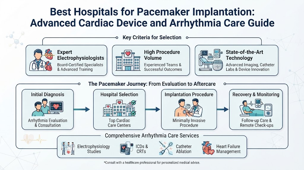
Selecting the right hospital for a cardiac procedure involves evaluating several structural and professional markers of quality care. Patients and families should focus on institutions that emphasize medical safety, clinical transparency, and structured follow-up.
Specialist Credentials and Electrophysiology Infrastructure
The single most important factor is the training and experience of the medical team. Patients should seek out hospitals that employ dedicated, board-certified cardiac electrophysiologists who possess extensive clinical experience in device implantation and rhythm management. Look for facilities that feature dedicated, state-of-the-art cardiac catheterization and electrophysiology labs that are exclusively utilized for rhythm procedures rather than general surgeries.
Emergency Infrastructure and Multidisciplinary Care
A premier cardiac facility must possess comprehensive emergency support. Even though pacemaker implantation is a highly standardized procedure, having 24/7 cardiac intensive care units (ICUs) and on-site cardiothoracic surgery teams provides an indispensable safety net should unexpected anatomical variations arise. Furthermore, top institutions utilize an integrated, multidisciplinary heart care team framework—where electrophysiologists, heart failure specialists, and specialized device nurses collaborate closely to manage all aspects of your cardiovascular health.
Comprehensive Long-Term Care Continuity
Because a pacemaker remains with you for life, the hospital’s operational follow-up infrastructure is vital. Confirm that the institution features a dedicated, structured device clinic that actively manages remote monitoring configurations, provides direct lines of communication for technical questions, and systematically coordinates long-term follow-up visits.
Hospital Selection Checklist
| Feature | Why It Matters | What to Look For |
| Electrophysiology Specialists | Ensures accurate diagnosis, optimal device selection, and safe placement | Board certifications, extensive procedure history, dedicated rhythm focus |
| Advanced Cath / EP Lab | Minimizes procedural risks through optimal visual and technical support | High-resolution low-dose fluoroscopy, digital electrical mapping systems |
| Modern Pacemaker Options | Provides access to customized traditional and leadless pacing styles | Availability of leadless systems and specialized biventricular (CRT) models |
| Device Programming Services | Optimizes individual device performance and preserves battery longevity | In-house programming software, specialized pacing technicians on staff |
| Remote Monitoring Support | Delivers continuous clinical oversight from the comfort of your home | Established remote console setup, automated arrhythmia alert systems |
| Emergency Cardiac Support | Guarantees complete safety if unexpected anatomical challenges occur | On-site cardiothoracic surgeons, 24/7 cardiac intensive care infrastructure |
Preparing for Pacemaker Implantation
Proper pre-operative preparation ensures that the procedure can be performed safely and efficiently with minimal risk of avoidable complications.
Clinical History and Diagnostic Review
In the days leading up to the procedure, the cardiac care team conducts an exhaustive review of your complete medical history. You will undergo standard pre-operative blood tests to evaluate kidney function, check electrolyte balances, and assess blood clotting parameters. A baseline chest X-ray and a fresh 12-lead ECG are typically documented to serve as anatomical references for the implantation team.
Medication Management and Adjustments
One of the most critical preparatory steps involves coordinating your current medications. Your cardiologist will provide specific instructions regarding blood thinners (anticoagulants) or antiplatelet drugs. In many instances, these medications may need to be temporarily paused or adjusted for a few days before the procedure to minimize the risk of bleeding around the surgical pocket site. Specific instructions will also be provided for managing diabetes medications and blood pressure therapies on the morning of the procedure.
Final Pre-Procedure Actions and Informed Consent
Patients are required to fast (nothing to eat or drink) for a specified number of hours before the scheduled surgery, typically starting at midnight the evening before. Before moving into the electrophysiology suite, the clinical team will systematically discuss the procedural path, answer outstanding questions, and complete the formal informed consent process.
What Happens During the Procedure?
Pacemaker implantation is typically performed under local anesthesia inside a specialized cardiac catheterization or electrophysiology laboratory, maintaining a highly structured and safe clinical sequence.
[Hospital Admission & IV Setup] ➔ [Local Anesthesia & Sedation] ➔ [Incision & Venous Access] ➔ [Lead Placement & Testing] ➔ [Pulse Generator Connection] ➔ [Incision Closure & Observation]
Step 1: Admission and Local Sedation
Once admitted and positioned comfortably on the procedure table, an intravenous (IV) line is established to deliver fluids, mild relaxing sedatives, and preventative antibiotics. The surgical site—typically just below the collarbone on the non-dominant side—is carefully shaved, cleansed with a sterile antiseptic solution, and draped. A local anesthetic is injected to completely numb the area where the incision will be made.
Step 2: Incision and Venous Access
The electrophysiologist makes a small, horizontal incision (typically two to three inches long) in the skin just beneath the collarbone. Working carefully through this opening, they isolate a major vein that leads directly to the heart. A small pocket is then created beneath the skin and fat layer, or over the chest muscle, to house the pulse generator.
Step 3: Lead Placement and Device Testing
Using high-resolution, real-time fluoroscopy for visual guidance, the doctor carefully introduces the pacing leads into the vein and guides them into the appropriate heart chambers. Once the tips of the leads are securely anchored into the internal heart wall, they are connected to an external computer system. The team runs specialized diagnostic tests to verify that the leads can cleanly read the heart’s natural signals and stimulate the muscle using a minimal amount of battery energy.
Step 4: Connecting the Generator and Incision Closure
Once the electrical testing confirms optimal placement, the ends of the leads are securely screwed into the pulse generator. The generator capsule is then gently tucked into the prepared subcutaneous chest pocket. The doctor meticulously closes the surgical incision using layers of dissolvable sutures and applies a sterile, protective adhesive dressing.
Depending on the patient’s underlying condition, overall stability, and specific hospital protocols, many individuals are comfortably discharged the very same day, while others may stay for a routine overnight observation period to ensure complete structural stability.
Recovery and Living With a Pacemaker
Adapting to life with an implantable cardiac device is generally a smooth process, provided that patients follow a few straightforward post-operative guidelines during the initial healing phases.
Immediate Post-Operative Wound Care
Keeping the surgical wound completely clean and dry is vital to prevent localized infections. Patients should avoid showering or submerging the chest wound in water for the first several days, following the exact timeframe provided by their surgical team. The protective dressing should be left in place until it naturally peels away or is formally removed during your first post-operative checkup. Monitor the area daily for any swelling, warmth, drainage, or increasing redness.
Temporary Physical Activity Restrictions
To allow the newly placed leads to bind securely to the internal heart tissue, specific movement restrictions are recommended for the first four to six weeks:
- Arm Movements: Avoid raising the arm on the side of the device above shoulder height.
- Weight Limits: Refrain from lifting, pushing, or pulling heavy objects (typically anything over 10–15 pounds), including heavy grocery bags or luggage.
- Avoid Strenuous Exercise: Postpone vigorous activities like golf, tennis, swimming, or running until your electrophysiologist confirms that the leads are fully stabilized.
Long-Term Lifestyle and Environmental Precautions
Modern pacemakers are built with robust protective shielding against everyday electrical equipment. It is entirely safe to use household appliances like microwave ovens, televisions, computers, and standard smartphones. However, a few practical rules should be integrated into your routine:
- Smartphones: Avoid placing your smartphone directly over the device in a breast pocket. Hold the phone to the ear on the opposite side of your pacemaker.
- Security Scanners: When passing through airport or retail security checkpoints, walk through at a normal pace. Do not linger near the scanners. Always carry your official pacemaker identification card to present to security personnel if a manual wand search is required.
- Medical Environments: Always inform all medical professionals, including dentists and physical therapists, that you have an implanted device. Certain diagnostic therapies—most notably standard Magnetic Resonance Imaging (MRI) scans—can generate intense magnetic fields that interfere with device function. While many modern pacemakers are explicitly rated as “MRI-conditional,” the device must always be evaluated and temporarily programmed into a safe mode before entering an MRI suite.
Possible Risks and Complications
While serious complications are uncommon and significantly reduced through experienced cardiac care teams and regular follow-up, it is important to understand the potential risks associated with any surgical procedure.
- Localized Bleeding or Hematoma: A collection of blood can sometimes form within the surgical pocket beneath the collarbone during the initial post-operative hours. This is carefully monitored and typically resolves naturally over several weeks.
- Incision Site Infection: Despite strict sterile techniques, bacteria can occasionally infect the skin wound or the deeper device pocket, presenting as localized swelling, drainage, or a fever. This requires prompt evaluation and antibiotic treatment.
- Lead Displacement: In a small percentage of cases, a pacing lead can shift or become dislodged from its anchor point in the heart wall during the early weeks of recovery. This displacement can cause the device to lose connection, occasionally requiring a brief revision procedure to reposition the wire.
- Pneumothorax: Because the veins used to access the heart lie very close to the upper lungs, a microscopic puncture of the lung membrane can occasionally occur during vein access, causing air to collect in the chest cavity. Most minor air pockets heal on their own, while larger ones are safely managed using a temporary chest tube.
- Device Malfunction: Mechanical or software failure within modern pulse generators is rare due to rigorous manufacturing standards and multi-layered backup systems.
- Future Battery Depletion: The pacemaker battery is designed to last for several years, depending on how frequently the device active paces the heart. When the battery runs low, the generator is easily replaced in a simple outpatient procedure.
How BESTCARDIACHOSPITALS Helps Patients
Navigating complex cardiovascular treatment paths can feel overwhelming for patients and families. BESTCARDIACHOSPITALS serves as an objective, educational platform designed to streamline your search for high-quality cardiac care.
Verified Hospital and Specialist Discovery
The platform provides access to a comprehensive directory of medical centers featuring specialized cardiac electrophysiology departments. Users can explore facility locations, evaluate operational infrastructure, and review the medical credentials of practicing electrophysiologists without needing to sort through unverified online sources.
Transparent Care Comparison and Educational Libraries
Through easy-to-read guides and comprehensive facility overviews, the platform allows you to evaluate cardiac centers based on objective quality markers, such as international healthcare accreditations, procedural safety track records, and the availability of advanced pacing technologies like leadless systems. This helps clarify complex medical terms, ensuring you know exactly what options to discuss with your doctor.
Direct Access to Practical Information
Beyond serving as a trusted educational resource, the platform allows users to easily submit requests for general treatment information and facility quotes. By facilitating communication between families and accredited healthcare systems, the platform helps you manage the logistical and financial planning of your cardiac care journey smoothly and confidently.
Common Mistakes People Make
Avoiding common behavioral errors after being recommended for pacing therapy can protect you from preventable complications and ensure a smoother recovery.
Delaying Treatment for Symptomatic Bradycardia
Ignoring warning signs like recurrent fainting spells, chronic dizziness, or extreme fatigue because of a fear of surgery is a serious mistake. A severely slow heart rhythm can lead to sudden falls, physical injury, or worsening heart strain if left unaddressed.
Missing Scheduled Follow-Up and Device Appts
Assuming that your pacemaker requires no further oversight once the surgical incision has healed is a major misconception. Skipping follow-up visits prevents your care team from checking battery usage, verifying lead integrity, and fine-tuning your programming settings.
Neglecting Early Post-Operative Wound Care
Washing the chest incision too early, removing protective dressings aggressively, or touching the wound with unclean hands elevates your risk of localized bacterial infections. Following your surgical team’s exact hygiene instructions is essential.
Forgetting to Carry Device Identification
Failing to keep your official pacemaker identification card in your wallet or phone case can cause unnecessary delays and complications at airport security checkpoints or during unexpected medical emergencies. Always keep this documentation with you.
Skipping Routine Device Checks and Updates
Even if you feel completely healthy, skipping regular clinic visits or failing to keep your home remote monitoring console plugged in stops your electrophysiologist from receiving vital diagnostic data that protects your long-term safety.
Common Myths vs. Facts
Myth: An implanted pacemaker completely replaces the natural physical heart.
Fact: A pacemaker does not replace the heart muscle. It acts as an electronic support system, working alongside your natural heart to monitor and correct electrical rhythm disruptions only when needed.
Myth: People with pacemakers must lead sedentary lives and avoid all physical exercise.
Fact: The vast majority of patients return to their normal daily activities and enjoy low-impact exercise routines like walking, swimming, and cycling once the initial recovery period is complete.
Myth: Every pacemaker device utilized in modern hospitals is exactly the same.
Fact: Pacemakers are highly customized. Devices range from single-chamber models to dual-chamber, biventricular systems, and leadless capsules, with each selection tailored to the patient’s specific heart condition.
Myth: Once an implantable pacemaker is in place, it never requires any form of clinical follow-up.
Fact: Consistent long-term monitoring is essential. Regular evaluations—whether in-person or via remote telemetry transmitters—are required to verify battery life, assess lead performance, and adjust settings.
Real-Life Educational Scenarios
Scenario A: Restoring Energy After Symptomatic Bradycardia
An individual experienced frequent dizziness and struggled to walk short distances due to a resting heart rate that consistently dropped to 42 beats per minute. Following a comprehensive electrophysiology evaluation, they received a dual-chamber pacemaker. The device safely stabilized their heart rate, completely resolving their dizzy spells and restoring their physical energy levels.
Scenario B: Overcoming Heart Block
A patient was diagnosed with a high-degree heart block after collapsing unexpectedly at home. They were admitted to a specialized cardiac hospital, where a dedicated team implanted a biventricular pacemaker. The synchronized pacing successfully restored proper coordination between the upper and lower heart chambers, significantly improving the patient’s daily quality of life.
Scenario C: Transitioning to Remote Monitoring
A patient living in a rural community required routine checks for their cardiac device but found regular long-distance travel difficult. Their cardiac center provided a wireless home remote monitoring console. The console securely transmitted device diagnostic data to the hospital while the patient slept, reducing the need for frequent travel while ensuring continuous clinical oversight.
Scenario D: Selecting a Specialized Treatment Center
A family sought advanced care options for an elderly relative who needed a pacemaker but also had complex underlying health conditions. By using educational resources to compare cardiac facilities, they selected a hospital featuring digital electrophysiology labs and a multidisciplinary heart failure team. The specialized team successfully managed the patient’s care pathway, providing a safe procedure and a well-coordinated recovery plan.
FAQs (Frequently Asked Questions)
1.What is a pacemaker?
A pacemaker is a small, surgically implanted medical device that monitors the heart’s natural electrical rhythm and delivers gentle electrical impulses to correct slow or irregular heartbeats.
2.Why is a pacemaker recommended?
It is typically prescribed to treat symptomatic bradycardia (a very slow heart rate), heart blocks (disrupted internal signaling), sick sinus syndrome, and certain forms of heart failure.
3.Who performs the pacemaker implantation surgery?
The procedure is performed by a cardiac electrophysiologist—a specialized cardiologist who focus exclusively on diagnosing and treating heart rhythm disorders.
4.How long does the implantation procedure take?
A standard pacemaker implantation typically takes between one to two hours to complete, depending on the number of pacing leads required and the patient’s individual anatomy.
5.Is the pacemaker implantation procedure painful?
No. The procedure is performed under local anesthesia to completely numb the upper chest area, combined with mild intravenous sedation to ensure the patient remains relaxed and comfortable throughout.
6.What is a leadless pacemaker?
A leadless pacemaker is a self-contained capsule inserted via a leg vein directly into the heart, completely eliminating the need for surgical chest incisions and pacing wires.
7.How long does initial recovery take?
Most patients return to light daily activities within a few days. However, specific restrictions—such as avoiding heavy lifting or raising the arm above the shoulder—should be maintained for four to six weeks.
8.Can I exercise after getting a pacemaker?
Yes. After the initial healing phase is complete and your doctor confirms the leads are stable, most individuals can safely return to regular, low-impact exercise like walking or cycling.
9.How often does the device need to be checked?
Devices are typically evaluated every three to six months. This oversight is often managed through a combination of automated home remote monitoring and annual in-person clinic visits.
10.What main environmental precautions should I follow?
Avoid placing smartphones directly over the device, walk through security scanners at a normal pace, and always inform medical teams that you have a pacemaker before undergoing specialized therapies like MRIs.
11.Can a pacemaker battery be replaced?
Yes. The battery inside the pulse generator lasts for several years. When it runs low, the generator is easily replaced in a straightforward outpatient procedure using the existing leads.
12.What are the main risks of the procedure?
Potential risks include localized bleeding or a bruise at the surgical site, infection of the wound pocket, shifting of a lead wire, or a minor pocket of air outside the lung.
13.How do I choose the right cardiac hospital?
Look for accredited medical centers that feature dedicated electrophysiology labs, employ experienced specialists, offer comprehensive remote monitoring programs, and provide 24/7 emergency cardiac care.
14.What technologies improve modern pacemaker care?
Key advancements include miniaturized leadless pacing systems, high-resolution real-time fluoroscopy, wireless home telemetry consoles, and computerized device programming software.
15.How does BESTCARDIACHOSPITALS help patients find care?
The platform provides a comprehensive directory of verified cardiac hospitals, outlines specialist credentials, shares educational heart resources, and simplifies the process of requesting treatment information.
Final Thoughts
Addressing heart rhythm disorders through timely medical intervention is an essential step toward protecting your cardiovascular health and maintaining your daily energy levels. Pacemaker implantation remains a highly dependable, standardized care pathway that offers profound benefits when managed by experienced cardiac electrophysiology teams. By selecting specialized hospitals equipped with modern digital laboratories and comprehensive follow-up programs, you ensure that every stage of your care—from the initial evaluation through long-term monitoring—is guided by clinical expertise and rigorous safety standards.