Introduction
Heart health is a cornerstone of overall well-being, and having access to precise diagnostic tools is essential for maintaining a healthy cardiovascular system. Among these tools, the echocardiogram stands out as one of the most versatile and informative procedures in modern medicine. By providing real-time, detailed images of the heart’s structure and function, this test helps doctors make informed decisions about your care.In this guide, we will explore the role of echocardiography, the different types of heart imaging available, and how specialized centers play a vital role in early detection. For those seeking reliable information on identifying high-quality cardiac centers, comparing diagnostic services, and learning more about cardiovascular care, BestCardiacHospitals offers a valuable resource to support your heart health journey.
What Is Echocardiography?
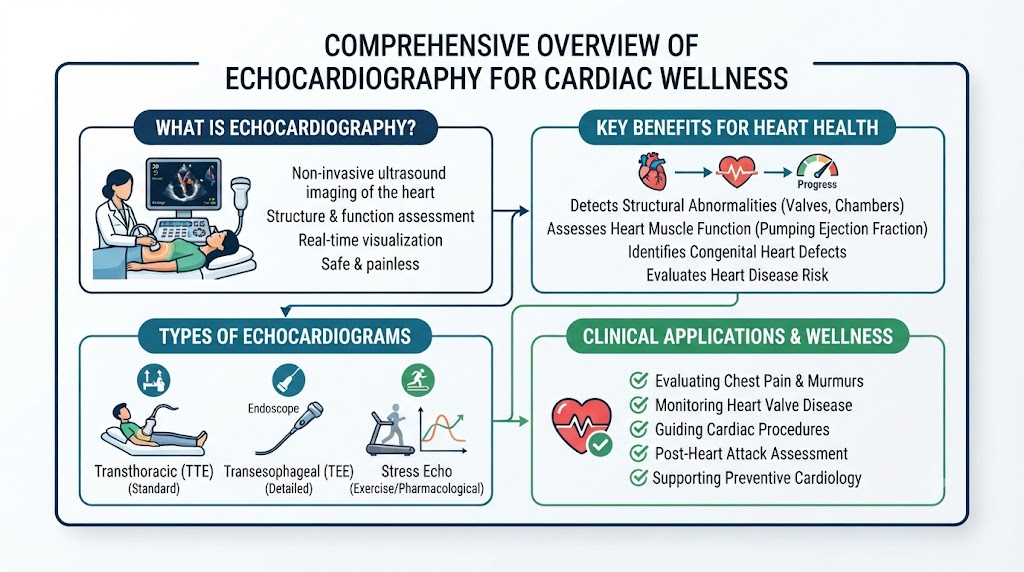
Echocardiography is a specialized, non-invasive diagnostic test that uses sound waves to create moving images of your heart. Often referred to as a “heart ultrasound,” this procedure is completely painless and does not use radiation.
How It Works
During the test, a technician moves a small device called a transducer over your chest. This device emits high-frequency sound waves that bounce off your heart’s structures—such as valves, walls, and chambers—and return to the machine. These echoes are converted into real-time images on a monitor.
Echocardiography vs. ECG
While both are vital, they serve different purposes. An Electrocardiogram (ECG) records the electrical activity of the heart to detect rhythm abnormalities, whereas an echocardiogram focuses on the physical structure and functional movement of the heart.
Why Specialized Cardiac Centers Matter
Choosing a center that specializes in cardiology can significantly impact the quality of your diagnostic care. Specialized facilities offer:
- Experienced Cardiologists: Experts who specialize in interpreting complex imaging data to provide accurate assessments.
- Advanced Imaging Departments: Access to the latest ultrasound machines that offer superior clarity and detail.
- Integrated Care: If an abnormality is detected, these centers often provide seamless transitions to treatment planning and specialist consultations.
- Emergency Support: Specialized centers are equipped to handle urgent cardiac diagnostic needs, ensuring immediate care when every minute counts.
Types of Echocardiography
Different situations require different views of the heart. Your cardiologist will recommend the best approach based on your symptoms and clinical history.
Echocardiography Types Comparison Table
| Test Type | Purpose | Common Uses |
| Transthoracic Echo (TTE) | Standard heart imaging | General heart evaluation |
| Transesophageal Echo (TEE) | Detailed heart images | Valve disorders and blood clots |
| Stress Echo | Heart function during exercise | Coronary artery disease assessment |
| Doppler Echo | Blood flow analysis | Valve function and circulation |
| 3D Echo | Advanced heart imaging | Surgical planning/structural assessment |
Conditions Diagnosed with Echocardiography
Echocardiography is a powerful tool for identifying a wide range of cardiovascular issues, including:
- Heart Valve Disorders: Detecting narrowing or leaking valves.
- Heart Failure: Assessing how well the heart pumps blood to the rest of the body.
- Congenital Heart Defects: Identifying structural issues present from birth.
- Cardiomyopathy: Evaluating changes in the heart muscle structure.
- Heart Infections: Identifying abnormalities like endocarditis.
Common Symptoms That May Require an Echocardiogram
Doctors often order an echocardiogram when patients present with persistent symptoms that suggest heart strain, such as:
- Unexplained chest pain or pressure.
- Shortness of breath, especially during physical activity.
- Sensations of palpitations or an irregular heartbeat.
- Recurrent dizziness, fainting, or severe fatigue.
- Swelling in the legs, ankles, or abdomen.
The Diagnostic Process
Before your procedure, your medical team will review your health history and perform a physical exam. You may also undergo an ECG or blood pressure check to provide a complete picture of your heart’s health.
Diagnostic Tests Comparison Table
| Test | Purpose | Benefit |
| ECG | Measures electrical activity | Detects rhythm abnormalities |
| Echocardiography | Heart structure and function | Detailed heart imaging |
| Stress Test | Evaluates exercise response | Detects reduced blood flow |
| Cardiac CT | Detailed anatomical imaging | Assesses coronary arteries |
Advanced Technologies Used in Cardiac Imaging Centers
The field of cardiac diagnostics is constantly evolving. Leading centers leverage advanced technologies to improve the precision of their findings:
- Digital 2D Echocardiography: The foundation of standard cardiac imaging.
- 3D/4D Echocardiography: Provides depth and volume, essential for planning complex cardiac procedures.
- Contrast Echocardiography: Uses safe contrast agents to enhance the visibility of the heart’s chambers.
- AI-Assisted Imaging: Helps cardiologists analyze data more efficiently and accurately.
How to Choose the Right Cardiac Center
When selecting a facility for your diagnostic needs, consider the following checklist:
- Experienced Cardiologists: Ensure the team has specialized credentials in imaging.
- Accreditation: Look for centers accredited for quality and safety in cardiac testing.
- Modern Equipment: Verify that the center uses updated, high-resolution ultrasound systems.
- Integrated Services: Does the center offer follow-up appointments and clear communication with your primary doctor?
Benefits of Early Heart Imaging
Early detection is often the key to successful management. Imaging can reveal subtle changes in heart function long before symptoms become severe, allowing for:
- Timely interventions.
- Better monitoring of conditions like high blood pressure.
- A clearer roadmap for preventive care and lifestyle adjustments.
Myths vs. Facts
| Myth | Fact |
| Echocardiography uses radiation | It uses safe, high-frequency sound waves |
| The test is painful | It is non-invasive and generally painless |
| Only older adults need heart imaging | People of all ages may need it based on symptoms |
| It replaces all other tests | It is part of a broader diagnostic approach |
Real-Life Patient Scenarios
- Early Detection: A patient with mild fatigue undergoes an echo, which reveals a treatable valve condition before it progresses to heart failure.
- Monitoring: A patient with a known heart condition uses regular, scheduled imaging to ensure their medication is working effectively.
- Precision Diagnosis: A patient with chest pain during exercise undergoes a stress echo, allowing doctors to identify the exact cause and start targeted therapy.
FAQs
- What is an echocardiogram?
A safe, non-invasive ultrasound of the heart. - Is echocardiography painful?
No, it is a comfortable, non-invasive procedure. - How long does it take?
Most standard echoes take between 30 to 60 minutes. - Why recommend an echo?
To assess heart structure, valve function, and pumping strength. - Difference between ECG and Echo?
ECG checks electricity; Echo checks physical structure. - Do I need to prepare?
Most tests require no special preparation, but follow specific instructions if you are having a stress or TEE test. - Is it safe during pregnancy?
Yes, it uses no radiation and is considered safe. - What can it detect?
Valve disease, heart failure, muscle weakness, and more. - How often should I have one?
This is determined by your cardiologist based on your medical history. - What happens after?
You can usually return to your normal daily activities immediately. - Is Doppler different?
It is a feature often used during an echo to measure blood flow velocity. - Can it detect heart failure?
Yes, it is the standard test for evaluating heart function. - Is fasting required?
Only for specific types, like the TEE test. - How do I choose a center?
Look for accredited labs, expert staff, and modern equipment. - When should I see a cardiologist?
If you have heart-related symptoms, risk factors, or concerning test results.
Final Thoughts
Echocardiography is a vital tool in modern medicine, providing the clarity needed to protect heart health. By choosing specialized diagnostic centers, you ensure that your heart health is in the hands of experts equipped with the latest technology. We encourage anyone experiencing heart-related symptoms or those with underlying risk factors to consult a qualified cardiologist. For further guidance, hospital comparisons, and reliable information regarding cardiac diagnostic services, explore the resources at BestCardiacHospitals.