Introduction
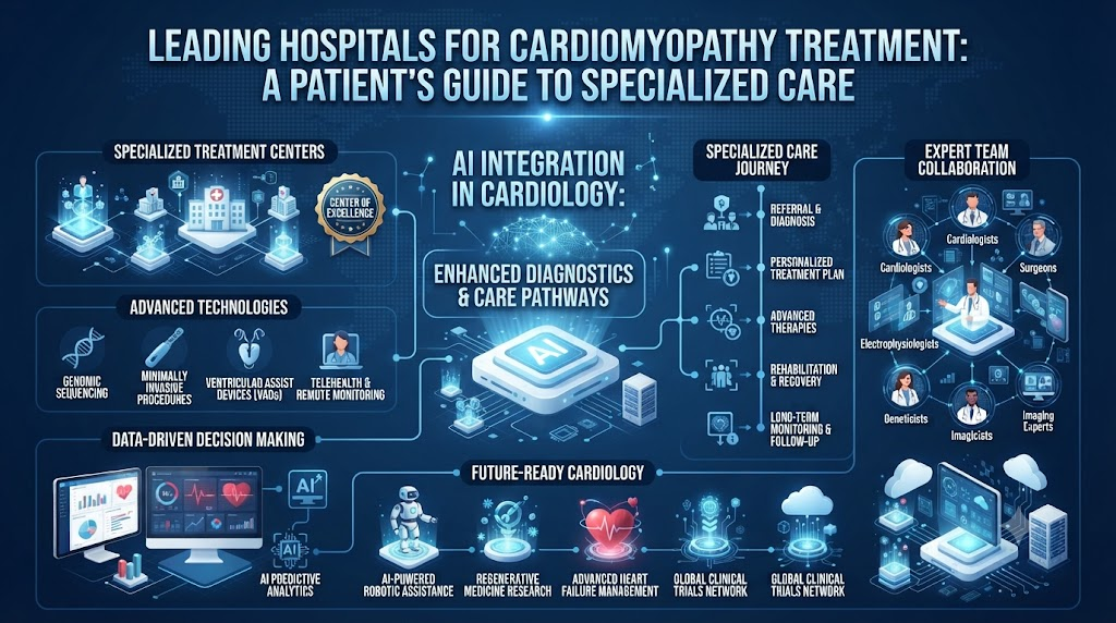
The heart muscle serves as the primary engine of the cardiovascular system, continuously contracting to pump oxygen-rich blood throughout the entire body. When a condition directly impacts the structure and function of this vital tissue, it can significantly alter a person’s overall health and quality of life. Cardiomyopathy refers to a group of diseases that affect the heart muscle, making it harder for the heart to pump blood effectively to the rest of the body.Selecting a medical center for cardiovascular care is a significant decision. Evaluating hospitals based on their diagnostic capabilities, multidisciplinary cardiology teams, electrophysiology services, and heart failure care models ensures patients and families receive care tailored to their condition. Platforms like BESTCARDIACHOSPITALS help readers understand cardiac conditions, learn about specialized hospital services, and compare available care options to support informed decision-making.
What Is Cardiomyopathy?
Cardiomyopathy is a progressive disease of the myocardium (heart muscle). Under healthy conditions, the heart’s muscular walls contract and relax in a coordinated rhythm, maintaining adequate blood flow and pressure. In cardiomyopathy, the heart muscle becomes abnormally enlarged, thickened, rigid, or replaced by scar tissue.
HEALTHY HEART                           CARDIOMYOPATHY
┌─────────────────────────┐             ┌─────────────────────────┐
│ • Normal muscle wall    │             │ • Thickened/Stiff wall  │
│   thickness             │     VS.     │ • Stretched chambers    │
│ • Efficient contraction │             │ • Reduced pumping/      │
│ • Normal blood filling  │             │   filling efficiency    │
└─────────────────────────┘             └─────────────────────────┘
These structural changes impair the heart’s functional capabilities in two main ways:
- Systolic Dysfunction: The heart muscle weakens and cannot pump blood out to the body with sufficient force.
- Diastolic Dysfunction: The heart muscle becomes stiff or rigid, preventing the ventricles (lower heart chambers) from relaxing and filling with blood properly between beats.
Cardiomyopathy can be inherited (passed down through family genetics) or acquired (developing as a result of other cardiovascular conditions, infections, metabolic disorders, or environmental factors). Depending on the cause and progression, some individuals remain asymptomatic for long periods, while others experience progressive heart failure, fluid retention, or abnormal heart rhythms (arrhythmias).
Types of Cardiomyopathy
Because cardiomyopathy presents in various structural forms, proper classification is essential for guiding treatment strategies.
Dilated Cardiomyopathy (DCM)
Dilated Cardiomyopathy is the most common form of heart muscle disease. It occurs when the heart’s main pumping chamber—the left ventricle—becomes enlarged (dilated) and stretched thin. As the chamber stretches, the muscle weakens and loses its ability to pump blood efficiently, often leading to heart failure.
- Possible Causes: Ischemic heart disease (prior heart attacks), high blood pressure, viral infections (myocarditis), heavy alcohol consumption, exposure to certain toxins or chemotherapy agents, and genetic gene mutations.
Hypertrophic Cardiomyopathy (HCM)
Hypertrophic Cardiomyopathy is characterized by abnormal thickening (hypertrophy) of the heart muscle walls, most commonly affecting the septum that divides the left and right ventricles. The thickened muscle can restrict the space inside the ventricles and, in some cases, physically block blood flow out of the heart (obstructive HCM).
- Inherited Component: HCM is frequently a genetic condition caused by mutations in heart muscle proteins. It can affect people of any age, including young athletes, and requires specialized screening for blood relatives.
Restrictive Cardiomyopathy (RCM)
Restrictive Cardiomyopathy is a less common form where the heart muscle walls become rigid, stiff, and unyielding, though the muscle thickness itself may remain normal. While the heart may pump normally during contractions, it cannot relax adequately between beats, restricting blood from filling the chambers properly.
- Possible Causes: Systemic infiltrate disorders such as amyloidosis (abnormal protein accumulation), sarcoidosis (inflammatory cell clusters), hemochromatosis (iron overload), or scarring following radiation therapy.
Arrhythmogenic Cardiomyopathy (ACM)
Formerly known as Arrhythmogenic Right Ventricular Cardiomyopathy (ARVC), this complex form occurs when normal heart muscle tissue is progressively replaced by fatty and fibrous scar tissue. This disruption impairs structural pumping and interferes with the heart’s electrical pathways, increasing the risk of dangerous ventricular arrhythmias.
Takotsubo Cardiomyopathy (Stress Cardiomyopathy)
Often called “broken heart syndrome,” Takotsubo cardiomyopathy involves a sudden, temporary weakening of the heart muscle (typically the apex of the left ventricle). It is frequently triggered by severe physical or emotional stress, leading to symptoms that closely mimic a acute heart attack. With prompt medical support, heart function often returns to baseline over several weeks.
Symptoms of Cardiomyopathy
In the early stages of cardiomyopathy, many individuals experience no noticeable symptoms. However, as the structural changes progress and the heart’s pumping or filling capacity declines, symptoms typically develop gradually:
- Shortness of Breath (Dyspnea): Breathlessness during routine physical activities, when lying flat in bed (orthopnea), or waking up suddenly at night gasping for air.
- Fatigue and Weakness: Persistent tiredness due to reduced oxygenated blood delivery to skeletal muscles and vital organs.
- Fluid Retention (Edema): Swelling in the lower legs, ankles, feet, abdomen, or neck veins caused by fluid accumulation.
- Chest Discomfort or Pressure: Pain, tightness, or pressure, particularly following physical exertion or heavy meals.
- Heart Palpitations: Sensations of a rapid, fluttering, pounding, or irregular heartbeat.
- Dizziness and Lightheadedness: Feeling unsteady or faint, often due to temporary drops in blood pressure or cardiac output.
- Fainting (Syncope): Sudden loss of consciousness, which can be an important warning sign of an underlying cardiac rhythm disturbance.
- Reduced Exercise Capacity: Decreased ability to keep up with previously manageable physical tasks, workouts, or daily walking.
Cardiomyopathy Risk Factors and Causes
Cardiomyopathy can result from genetic factors, underlying medical conditions, or environmental exposures. Identifying the root cause helps physicians tailor treatment strategies effectively.
- Family History and Genetics: Many forms of cardiomyopathy—particularly Hypertrophic Cardiomyopathy and Arrhythmogenic Cardiomyopathy—have a strong genetic origin. A family history of unexplained heart failure, enlarged heart, or sudden cardiac death indicates a higher risk.
- Chronic High Blood Pressure (Hypertension): Uncontrolled high blood pressure forces the heart muscle to work harder to pump blood, causing the muscle walls to thicken, stiffen, and eventually weaken over time.
- Coronary Artery Disease & Heart Attacks: Blocked coronary arteries reduce blood supply to the heart tissue. A prior heart attack leaves scar tissue, impairing the heart muscle’s ability to contract normally (ischemic cardiomyopathy).
- Viral and Inflammatory Conditions: Viral infections (such as enteroviruses, adenovirus, or COVID-19) can cause myocarditis (inflammation of the heart muscle), which may lead to dilated cardiomyopathy.
- Medications, Alcohol, and Toxins: Prolonged heavy alcohol consumption, illicit drug use (such as cocaine or amphetamines), and certain cancer treatments (such as anthracycline chemotherapy agents or radiation therapy to the chest) can cause damage to cardiac tissue.
- Metabolic and Systemic Disorders: Diabetes, thyroid imbalances, chronic kidney disease, auto-immune conditions (such as lupus), and infiltrative diseases (like amyloidosis or sarcoidosis) can impair heart muscle function.
- Pregnancy-Related (Peripartum Cardiomyopathy): A rare form of heart failure that develops during the final month of pregnancy or within the first five months following delivery, causing the heart chambers to dilate and weaken.
How Cardiomyopathy Is Diagnosed
Accurate diagnosis requires evaluating the heart’s physical structure, pumping efficiency, valve function, and electrical activity. According to clinical guidance from the National Heart, Lung, and Blood Institute (NHLBI) and the National Institutes of Health (NIH), non-invasive imaging forms the core of initial assessment, with genetic evaluations added in specific clinical scenarios.
DIAGNOSTIC WORKUP FOR CARDIOMYOPATHY
┌──────────────────┐    ┌──────────────────┐    ┌──────────────────┐
│  INITIAL TESTS   │ ──►│ CARDIA IMAGING   │ ──►│ ADVANCED WORKUP  │
│ • Medical History│    │ • Echocardiogram │    │ • Cardiac MRI    │
│ • Physical Exam  │    │ • ECG / Holter   │    │ • Genetic Testing│
│ • Blood Markers  │    │ • Chest X-Ray    │    │ • Cath / Biopsy  │
└──────────────────┘    └──────────────────┘    └──────────────────┘
- Medical History & Physical Examination: Evaluating personal symptoms, lifestyle factors, medication history, and detailed family history of cardiac conditions.
- Electrocardiogram (ECG / EKG): Records the electrical activity of the heart to detect abnormal rhythms, enlargement of heart chambers, or signs of prior muscle damage.
- Echocardiography (Transthoracic Echo): A fundamental diagnostic tool that uses ultrasound waves to produce real-time images of the heart. It allows cardiologists to measure chamber size, wall thickness, valve function, and ejection fraction (the percentage of blood pumped out of the main chamber with each beat).
- Cardiac Magnetic Resonance Imaging (Cardiac MRI): Provides detailed, high-resolution 3D images of heart tissue. It is particularly effective for identifying tissue scarring, fatty replacement (in ACM), or protein deposits (in amyloidosis).
- Holter and Rhythm Monitoring: Portable ECG devices worn for 24 hours to several weeks to detect intermittent arrhythmias or dangerous rhythm disruptions during daily activities.
- Blood Tests: Evaluating heart failure biomarkers (such as NT-proBNP or BNP), kidney and liver function, thyroid levels, inflammatory markers, and iron studies.
- Genetic Testing: Recommended for individuals diagnosed with inherited forms of cardiomyopathy (such as HCM or ACM) to identify specific gene mutations. Screening family members helps identify individuals at risk before symptoms appear.
- Cardiac Catheterization & Endomyocardial Biopsy: Invasive procedures where a thin catheter is guided through blood vessels into the heart to measure internal pressures directly, check for coronary artery blockages, or take small muscle tissue samples for analysis.
Cardiomyopathy Treatment Options
Treatment for cardiomyopathy aims to manage symptoms, slow disease progression, lower the risk of complications (such as heart failure or arrhythmias), and improve quality of life. Treatment plans are tailored to the individual based on the type, severity, and underlying cause of the condition.
Medications
Pharmacological therapy is the primary treatment for managing most forms of cardiomyopathy. Depending on the patient’s presentation, a cardiologist may prescribe:
- ACE Inhibitors, ARBs, or ARNI: Help relax blood vessels, lower blood pressure, and reduce workload on the heart.
- Beta-Blockers: Slow heart rate and lower blood pressure, protecting the heart muscle from stress hormones and reducing arrhythmia risks.
- Diuretics (Water Pills): Help the body eliminate excess fluid and sodium, reducing swelling in the legs and easing fluid congestion in the lungs.
- Mineralocorticoid Receptor Antagonists (MRAs): Help regulate fluid balance and prevent progressive scarring (fibrosis) of heart tissue.
- SGLT2 Inhibitors: Medications originally developed for diabetes that have been shown to improve outcomes and reduce hospitalizations in heart failure.
- Antiarrhythmics: Medications to help stabilize and control irregular heart rhythms.
- Blood Thinners (Anticoagulants): Reduce the risk of blood clots forming inside enlarged chambers or during irregular heart rhythms like atrial fibrillation.
Lifestyle Adjustments and Supportive Care
Non-pharmacological strategies support cardiac health and help manage day-to-day symptoms:
- Sodium and Fluid Regulation: Managing daily salt intake helps reduce fluid retention and maintain balanced blood pressure.
- Guided Physical Activity: Enrolling in a structured cardiac rehabilitation program provides supervised exercise tailored to individual physical limits.
- Avoiding Substances: Limiting or avoiding alcohol and avoiding tobacco products protects heart muscle function.
- Monitoring Body Weight: Daily weight monitoring helps detect sudden fluid accumulation early, allowing for timely medication adjustments.
Implantable Devices
For patients at risk of dangerous heart rhythms or progressive heart failure, specialized cardiac devices may be recommended:
- Implantable Cardioverter-Defibrillator (ICD): A small device implanted under the skin that continuously monitors heart rhythm. If a dangerous, life-threatening ventricular rhythm is detected, it delivers a precise electrical shock to restore a normal rhythm.
- Pacemaker: Delivers small electrical impulses to prevent the heart from beating too slowly.
- Cardiac Resynchronization Therapy (CRT): A specialized pacemaker (often combined with an ICD as a CRT-D) that coordinates the electrical signals sent to both ventricles, helping a weakened heart pump more synchronously and efficiently.
Surgical and Catheter-Based Procedures
When medications and devices are insufficient, structural interventions may be considered:
- Surgical Septal Myectomy: A specialized open-heart procedure for obstructive Hypertrophic Cardiomyopathy where a surgeon removes a small portion of the thickened septum, relieving blood flow obstruction.
- Alcohol Septal Ablation: A catheter-based procedure that injects a small amount of alcohol into a target artery, reducing a small portion of thickened septum tissue to widen the blood flow pathway.
- Catheter Ablation: Uses radiofrequency heat or cryo-cold energy to target and eliminate small areas of heart tissue that generate abnormal electrical signals causing persistent arrhythmias.
Advanced Heart Failure Treatment
For severe, end-stage cardiomyopathy that no longer responds adequately to standard medical therapies, advanced treatments may be required. Guidance from the American Heart Association (AHA) emphasizes that these situations benefit from evaluation by a multidisciplinary advanced heart failure team:
- Left Ventricular Assist Device (LVAD): A mechanical circulatory support pump surgically implanted to help a weakened left ventricle pump blood throughout the body. It can be used as a temporary bridge to heart transplantation or as long-term therapy.
- Heart Transplantation: The surgical replacement of a failing heart with a healthy donor heart for eligible patients with severe, end-stage heart failure.
Cardiomyopathy Treatment Comparison Table
| Treatment Approach | Primary Clinical Goal | Key Considerations & Factors |
| Prescription Medications | Manage symptoms, lower blood pressure, reduce fluid retention, protect heart tissue | Primary treatment; requires ongoing adjustment based on symptoms and kidney function. |
| Lifestyle & Dietary Care | Reduce stress on the heart, prevent fluid buildup, maintain general health | Requires ongoing patient engagement; includes low-sodium diet and supervised exercise. |
| Implantable Devices (ICD/CRT) | Prevent sudden cardiac arrest, correct irregular rhythms, improve pumping sync | Selected based on arrhythmia risk, ejection fraction, and specific ECG findings. |
| Septal Myectomy / Ablation | Relieve physical blood flow obstruction in Hypertrophic Cardiomyopathy | Recommended for symptomatic obstructive HCM when medical therapy is insufficient. |
| Catheter Ablation | Eliminate pathways causing persistent irregular heart rhythms | Evaluated by electrophysiologists when medications cannot control arrhythmias. |
| LVAD (Assist Device) | Provide mechanical pumping support for severe heart failure | Considered for end-stage heart failure as a bridge to transplant or destination therapy. |
| Heart Transplantation | Replace a failing heart with a healthy donor organ | Reserved for eligible patients with advanced disease when other treatments are exhausted. |
What Makes a Leading Cardiomyopathy Hospital?
Managing heart muscle disease requires specialized expertise and comprehensive medical infrastructure. Patients evaluating hospitals for complex cardiac care should consider several key factors:
KEY PILLARS OF CARDIAC EXCELLENCE
┌────────────────────────────────────────────────────────┐
│ 1. Subspecialized Cardiology Teams                      │
│ 2. Advanced Non-Invasive Cardiac Imaging (Echo / MRI)  │
│ 3. Comprehensive Electrophysiology & Device Services   │
│ 4. Dedicated Cardiac ICUs & Emergency Infrastructure   │
│ 5. Advanced Heart Failure & Mechanical Support Options │
└────────────────────────────────────────────────────────┘
- Experienced Cardiology Specialists: Access to board-certified cardiologists with fellowship training in heart failure, cardiomyopathy management, adult congenital heart disease, and electrophysiology.
- Multidisciplinary Cardiac Care Teams: Integrated teams that bring together heart failure specialists, cardiac surgeons, electrophysiologists, interventional cardiologists, cardiac radiologists, genetic counselors, specialized nurses, and cardiac rehabilitation dietitians.
- Advanced Diagnostic Infrastructure: Modern imaging technologies, including 3D echocardiography, dedicated cardiac MRI scanners, PET/CT capabilities, and on-site genetic testing programs.
- Comprehensive Electrophysiology Services: Specialized laboratories equipped for complex mapping and ablation of arrhythmias, as well as expert device implantation (ICDs, pacemakers, CRT).
- Cardiac Surgery and Structural Interventions: On-site cardiac surgical suites with experience in specialized procedures such as septal myectomy and valve repairs.
- Specialized Critical Care Units: Dedicated Cardiac Intensive Care Units (CICU) staffed 24/7 by critical care cardiologists and specialized nursing staff to support complex or unstable patients.
- Advanced Heart Failure & Transplant Capabilities: Hospitals offering mechanical circulatory support (LVAD) and heart transplantation provide a continuous spectrum of care for progressive conditions.
How to Compare Cardiomyopathy Hospitals
When evaluating cardiac care centers, patients and families should consider their specific medical needs rather than relying solely on generalized rankings:
- Specialist Expertise: Confirm that the hospital has dedicated heart failure and cardiomyopathy specialists who treat your specific condition regularly.
- Diagnostic Capabilities: Ensure the center offers high-resolution cardiac MRI, advanced echocardiography, and genetic counseling services.
- Treatment Capabilities: Verify that the hospital provides the full spectrum of care you may need, from medical management to electrophysiology and surgical procedures.
- Volume and Experience: Ask about the hospital’s annual volume for specific procedures, such as ICD implantations, catheter ablations, or septal myectomies.
- Emergency and ICU Infrastructure: Confirm the availability of a 24/7 cardiac ICU and emergency support services.
- Integrated Multidisciplinary Care: Check whether cardiologists, electrophysiologists, surgeons, and geneticists collaborate in regular care planning sessions.
- Patient Support and Rehabilitation: Ensure the facility offers structured cardiac rehabilitation, dietary counseling, and ongoing heart failure education.
- Financial Transparency: Request clear estimations regarding hospital admission, specialist consultations, diagnostic imaging, and procedure fees.
- Insurance Compatibility: Verify direct billing arrangements and insurance coverage with your healthcare provider.
- Accessibility and Follow-up Care: Consider travel distance and ease of access for routine follow-up consultations, medication monitoring, and device checks.
Cardiomyopathy Hospital Selection Checklist
| Factor to Evaluate | Key Elements to Verify | Patient Checklist Notes |
| Cardiology Team | Fellowship-trained heart failure and cardiomyopathy specialists | [ ] Confirmed specialist team |
| Diagnostic Tech | Availability of 3D Echocardiography, Cardiac MRI, and genetic testing | [ ] Advanced imaging available |
| Rhythm Care | Electrophysiology labs for ICD/pacemaker implantation and ablations | [ ] Electrophysiology service checked |
| Surgical Options | Cardiac surgical capabilities (septal myectomy, valve repair) | [ ] Surgical backup verified |
| Critical Care | 24/7 Dedicated Cardiac Intensive Care Unit (CICU) | [ ] Emergency / CICU checked |
| Advanced Therapies | Mechanical support (LVAD) and heart transplant programs | [ ] Advanced options reviewed |
| Follow-Up Care | Supervised cardiac rehabilitation and long-term outpatient monitoring | [ ] Rehab & follow-up confirmed |
| Cost & Insurance | Transparent pricing breakdown and direct insurance authorization | [ ] Insurance/Cost cleared |
Questions to Ask a Cardiologist
Preparing questions prior to your appointment helps ensure you understand your diagnosis, treatment plan, and long-term outlook:
- Diagnosis Details: What specific type of cardiomyopathy do I have, and what is its current severity?
- Underlying Cause: What is the likely cause of my condition, and is there any indication it could be genetic or inherited?
- Heart Function Assessment: What is my current ejection fraction, and what do my diagnostic imaging tests show regarding heart structure?
- Treatment Plan: What are my primary treatment options, and how will my medications help manage my symptoms?
- Specialist Referral: Would my condition benefit from evaluation by a heart failure specialist or electrophysiologist?
- Device Needs: Am I at risk for dangerous heart rhythms, and do I need an implantable cardioverter-defibrillator (ICD) or pacemaker?
- Family Screening: Should my immediate family members (children, siblings, parents) undergo genetic testing or screening echocardiograms?
- Lifestyle Guidelines: What physical activity levels, dietary sodium limits, and fluid intake goals should I follow?
- Warning Signs: What specific symptoms (such as sudden weight gain, increased swelling, or shortness of breath) should prompt an immediate call to your office?
- Follow-up Frequency: How often will I need follow-up appointments, blood work, and repeat diagnostic imaging?
When Cardiomyopathy May Require Specialized Care
While many patients manage cardiomyopathy through routine cardiology care, certain clinical situations warrant referral to a specialized heart failure center or advanced cardiac program:
- Progressive or Uncontrolled Symptoms: Experiencing worsening shortness of breath, severe fatigue, or persistent fluid retention despite taking prescribed medications.
- Declining Pumping Function: Experiencing a progressive drop in ejection fraction or worsening heart chamber dilation.
- Recurrent Hospital Admissions: Multiple emergency department visits or hospitalizations for heart failure stabilization within a short timeframe.
- Complex Rhythms or Fainting: Frequent episodes of heart racing, unexplained dizziness, or fainting spells (syncope).
- Suspected Inherited Conditions: A strong family history of early-onset heart failure, hypertrophic cardiomyopathy, or sudden unexplained cardiac death.
- Consideration for Advanced Therapies: Evaluating eligibility for specialized surgical interventions, implantable devices, LVAD support, or heart transplantation.
As highlighted in clinical guidelines from the American Heart Association, timely referral to an advanced heart failure team ensures complex cases receive comprehensive evaluation and access to the full range of treatment options.
Common Mistakes Patients Should Avoid
- Ignoring Early Symptoms: Dismissing gradual breathlessness or fatigue as normal aging or temporary tiredness instead of seeking medical evaluation.
- Stopping Prescribed Medications: Discontinuing heart medications independently when feeling better. Guidance from the American Heart Association stresses that heart medications should never be altered or stopped without consulting your cardiologist, as doing so can trigger sudden symptom flare-ups or complications.
- Overlooking Family History: Failing to share a family history of heart disease, enlarged heart, or sudden cardiac death with your physician.
- Choosing a Facility Based Only on Marketing: Selecting a hospital based solely on advertisements rather than verifying specialist expertise, diagnostic technology, and care volume.
- Skipping Follow-up Appointments: Missing routine cardiology visits, blood work, or diagnostic imaging, which are essential for monitoring disease progression and medication safety.
- Delaying Care After Fainting: Treating fainting episodes or sudden severe heart racing as minor events rather than seeking urgent evaluation for potential arrhythmias.
Myths vs Facts
| Myth | Fact |
| Myth: Cardiomyopathy is a single disease with one uniform treatment. | Fact: Cardiomyopathy includes several distinct types, each requiring a tailored diagnostic and treatment approach. |
| Myth: Every person diagnosed with cardiomyopathy will eventually need surgery. | Fact: Many individuals manage their condition effectively for years using medications, device therapy, and lifestyle modifications. |
| Myth: Cardiomyopathy always presents with obvious, severe symptoms. | Fact: In its early stages, cardiomyopathy can be subtle or asymptomatic, often discovered during routine medical exams. |
| Myth: Family history is irrelevant if you currently feel healthy. | Fact: Many forms of cardiomyopathy have genetic causes; screening family members can identify conditions before symptoms appear. |
| Myth: Physical activity should be completely avoided after diagnosis. | Fact: Most patients benefit from appropriate, medically supervised exercise; activity plans should be tailored with your cardiologist. |
| Myth: A hospital’s general reputation guarantees successful care for complex cases. | Fact: Specialized expertise in heart failure, advanced imaging, electrophysiology, and multidisciplinary planning are key to managing complex conditions. |
How BESTCARDIACHOSPITALS Helps Patients
Navigating cardiovascular healthcare options can feel overwhelming. BestCardiacHospitals.com provides educational resources to help patients and families understand cardiac conditions and evaluate care options effectively.
BESTCARDIACHOSPITALS PLATFORM ROLE:
┌─────────────────────────┐     ┌─────────────────────────┐     ┌─────────────────────────┐
│ LEARN ABOUT CONDITIONS  │ ──► │  EXPLORE CAPABILITIES   │ ──► │ COMPARE CARE OPTIONS    │
│ Understand types,       │     │ Research diagnostic     │     │ Evaluate multidisciplinary│
│ symptoms & diagnostics  │     │ and treatment services  │     │ teams and facilities    │
└─────────────────────────┘     └─────────────────────────┘     └─────────────────────────┘
The platform supports readers by:
- Explaining Complex Conditions: Providing clear explanations of cardiovascular diseases, diagnostic tests, and medical procedures.
- Outlining Hospital Services: Helping patients understand key hospital features, such as cardiac ICUs, electrophysiology labs, and specialized heart failure units.
- Offering Structural Frameworks: Supplying comparison criteria and evaluation checklists to help users compare healthcare options objectively.
- Supporting Patient Autonomy: Empowering individuals with practical questions and insights to prepare for discussions with their medical care team.
Frequently Asked Questions
1.What is cardiomyopathy?
Cardiomyopathy is a disease of the heart muscle that impairs its ability to pump blood effectively to the rest of the body, potentially leading to heart failure or arrhythmias.
2.What are the main types of cardiomyopathy?
The primary types include Dilated Cardiomyopathy (DCM), Hypertrophic Cardiomyopathy (HCM), Restrictive Cardiomyopathy (RCM), Arrhythmogenic Cardiomyopathy (ACM), and Takotsubo (stress) Cardiomyopathy.
3.What are common symptoms of cardiomyopathy?
Common symptoms include shortness of breath, persistent fatigue, swelling in the legs or ankles, heart palpitations, chest pressure, dizziness, and reduced exercise tolerance.
4.How is cardiomyopathy diagnosed?
Diagnosis typically involves medical history, physical examination, electrocardiogram (ECG), echocardiography, cardiac MRI, rhythm monitoring, and, in selected cases, genetic testing or cardiac catheterization.
5.Can cardiomyopathy be treated?
Yes. While many forms cannot be completely cured, cardiomyopathy can be managed using medications, lifestyle modifications, implantable devices (ICD/pacemaker), procedures, and advanced heart failure therapies.
6.What type of doctor treats cardiomyopathy?
Cardiomyopathy is treated primarily by cardiologists, often with input from specialized heart failure experts, electrophysiologists, and cardiac surgeons for complex cases.
7.Can cardiomyopathy cause heart failure?
Yes. Because cardiomyopathy weakens or stiffens the heart muscle, it is one of the leading underlying causes of chronic heart failure.
8.Do all cardiomyopathy patients need medication?
Most patients with symptomatic cardiomyopathy require prescription medications to help manage fluid balance, support pumping efficiency, lower blood pressure, or control heart rhythms.
9.When might an ICD be recommended?
An Implantable Cardioverter-Defibrillator (ICD) may be recommended for patients with significantly reduced ejection fraction or specific structural patterns that increase the risk of dangerous ventricular arrhythmias.
10.Can cardiomyopathy be inherited?
Yes. Several forms—especially Hypertrophic Cardiomyopathy and Arrhythmogenic Cardiomyopathy—are genetic conditions that can be passed down through families.
Conclusion
Cardiomyopathy encompasses a diverse group of heart muscle disorders that require accurate diagnosis and individualized treatment plans. Management strategies depend on the specific type, underlying cause, severity, and risk factors—ranging from daily lifestyle habits and medications to specialized cardiac devices and advanced heart failure care.When evaluating hospitals for treatment, patients should focus on centers with experienced cardiology teams, comprehensive diagnostic capabilities, multidisciplinary care models, and robust follow-up support. Understanding your condition and working closely with qualified cardiologists ensures a tailored approach to managing your cardiovascular health.