Introduction
Experiencing a complex heart rhythm disorder or being diagnosed with a high risk for sudden cardiac arrest can feel overwhelming for patients and their families. The human heart relies on a delicate electrical conduction system to contract predictably. When that electrical network encounters disruptions, dangerous irregularities—known as arrhythmias—can compromise blood flow to vital organs. For individuals facing severe ventricular arrhythmias, an Implantable Cardioverter Defibrillator (ICD) serves as a continuous, internal safeguard. Navigating these options empowers you to make proactive, well-informed choices for your cardiovascular health. To explore reliable educational resources and medical guidance on cardiac care, BESTCARDIACHOSPITALS offers comprehensive tools to help patients and families locate leading medical centers and better understand advanced heart treatments.
What Is an Implantable Cardioverter Defibrillator (ICD)?
An Implantable Cardioverter Defibrillator (ICD) is a small, battery-powered electronic device designed to monitor and treat abnormal heart rhythms (arrhythmias). Placed just beneath the skin—typically in the upper chest below the left collarbone—the device consists of a compact generator (housing the battery and computer circuitry) connected to one or more thin, flexible insulated wires called leads. These leads are threaded through veins directly into the heart’s chambers, where their exposed tips touch the heart tissue.
The primary function of an ICD is continuous vigilance. The internal computer processes every heartbeat, analyzing the rate, regularity, and electrical conduction patterns. If the heart maintains a normal, healthy rhythm, the ICD remains in a quiet monitoring state. However, if the device detects a life-threatening ventricular arrhythmia, it automatically executes pre-programmed therapy protocols designed to restore normal rhythm.
Heart Rhythm Monitoring
 ├── Normal Rhythm ──> Continuous Passive Surveillance
 └── Malignant Arrhythmia Detected
      ├── Ventricular Tachycardia ──> Anti-Tachycardia Pacing (ATP)
      └── Ventricular Fibrillation ──> High-Energy Defibrillation Shock
How the Device Works
An ICD delivers treatment using a tiered therapeutic approach depending on the speed and severity of the arrhythmia:
- Anti-Tachycardia Pacing (ATP): When the heart beats at an abnormally fast but regular rate (ventricular tachycardia), the ICD can issue a rapid series of low-energy electrical pulses. These subtle pulses interrupt the chaotic rhythm and restore normal beating without causing pain or discomfort to the patient.
- Cardioversion Therapy: If pacing pulses fail to stop the fast rhythm, or if the heart rate accelerates further, the device can deliver a synchronized, low-energy shock to reset the heart’s electrical system.
- Defibrillation Therapy: In cases of severe, chaotic heart rhythms (ventricular fibrillation) where the heart quivers uselessly instead of pumping blood, the ICD instantly delivers a higher-energy electrical shock. This shock momentarily stops all electrical activity, allowing the heart’s natural pacemaker to re-establish a stable rhythm.
- Bradycardia Pacing: If the heart rate drops dangerously low, the ICD can function like a standard pacemaker, sending gentle electrical impulses to keep the heart beating at a safe baseline rate.
Difference Between an ICD and a Pacemaker
Patients often confuse ICDs with conventional pacemakers because both are implantable cardiac devices that monitor heart rhythms. However, they serve distinct clinical purposes:
- Pacemaker: Primarily designed to treat slow heart rhythms (bradycardia). It continuously monitors the heart and sends mild, painless electrical signals whenever the heart rate drops too low.
- Implantable Cardioverter Defibrillator (ICD): Designed to treat fast, life-threatening heart rhythms (tachyarrhythmias) while also possessing the ability to pace for slow heart rates. Because an ICD contains high-voltage capacitors necessary to deliver high-energy shocks, the device generator is slightly larger than a traditional pacemaker.
Conditions Where ICD Therapy May Be Considered
Cardiologists evaluate ICD therapy for patients with specific cardiovascular conditions, including severe heart muscle weakness (cardiomyopathy), previous heart attacks that left significant scar tissue, inherited channelopathies, or a history of unexplained fainting accompanied by dangerous heart rhythms. Continuous monitoring ensures that if a sudden, dangerous electrical disruption occurs, life-saving treatment is administered within seconds.
Who May Need an ICD?
An ICD is not recommended for every individual with heart disease. Rather, clinical guidelines specify that ICD therapy is reserved for appropriately selected patients who have experienced a life-threatening arrhythmia or who meet specific risk criteria after a thorough cardiovascular evaluation.
Ventricular Tachycardia (VT)
Ventricular tachycardia is a rapid heart rhythm originating in the heart’s lower chambers (ventricles). Because the heart beats so quickly, its chambers do not have enough time to fill with blood between beats, leading to dizziness, shortness of breath, chest pain, or loss of consciousness. Sustained VT can rapidly deteriorate into ventricular fibrillation, making ICD implantation an important protective measure.
Ventricular Fibrillation (VF)
Ventricular fibrillation is an extreme medical emergency characterized by rapid, disorganized electrical signals that cause the ventricles to quiver uselessly. Blood flow to the brain and vital organs stops immediately, resulting in sudden cardiac arrest. An ICD provides immediate internal defibrillation if VF recurs.
Heart Failure with Reduced Ejection Fraction
Heart failure occurs when the heart muscle cannot pump blood efficiently throughout the body. Clinicians measure pumping efficiency using ejection fraction (EF)—the percentage of blood pumped out of the main reservoir with each contraction. A normal ejection fraction ranges between 50% and 70%. Patients with a significantly reduced ejection fraction (often 35% or lower) face an increased risk of sudden cardiac arrest, making primary prevention ICD evaluation an important component of their care.
Cardiomyopathy
Cardiomyopathy refers to diseases of the heart muscle that alter its structure and function. Whether dilated (stretched and thinned muscle), hypertrophic (abnormally thickened muscle), or arrhythmogenic (scar-replaced tissue), structural changes can disrupt normal electrical pathways and trigger severe ventricular arrhythmias.
Inherited Heart Rhythm Disorders
Certain genetic conditions affect the microscopic ion channels within heart cells without necessarily altering the physical structure of the heart. Conditions such as Long QT Syndrome, Brugada Syndrome, and Catecholaminergic Polymorphic Ventricular Tachycardia (CPVT) can cause sudden electrical instability. For selected high-risk patients with these conditions, an ICD provides an effective safety net.
Survivors of Sudden Cardiac Arrest
Individuals who have survived a sudden cardiac arrest caused by a ventricular arrhythmia undergo extensive clinical evaluation. When the underlying cause cannot be completely corrected through medication, surgery, or catheter intervention, secondary prevention guidelines typically recommend an ICD to protect against future events.
ICD Implantation Services Comparison Table
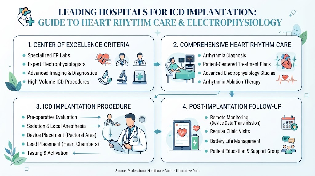
Understanding the different steps involved in ICD therapy helps patients navigate their care with confidence. Advanced heart centers structure their services into clear, connected stages:
| Service | Primary Purpose | Benefit to Patient |
| Comprehensive Cardiology Evaluation | Assesses overall heart function, ejection fraction, and clinical history. | Determines clear eligibility for device therapy based on guidelines. |
| Electrophysiology Consultation | Evaluates specific electrical pathways and arrhythmia patterns. | Provides an individualized treatment strategy tailored to your heart. |
| Precision ICD Implantation | Places leads and the generator using specialized imaging guidance. | Ensures safe, accurate device positioning with minimal discomfort. |
| Device Programming & Testing | Configures detection thresholds and therapeutic shock/pacing levels. | Tailors device responses to prevent unnecessary or inappropriate shocks. |
| Remote Device Monitoring | Wirelessly transmits device data from home to the care team. | Catches early signs of battery depletion, lead issues, or rhythm changes. |
| Long-Term Cardiac Follow-Up | Provides ongoing clinical reviews, medication adjustments, and support. | Maintains long-term heart stability and device functionality. |
Services Offered by Leading Hospitals
When evaluating medical facilities, it is helpful to look for hospitals that offer complete, end-to-end cardiovascular care. Advanced heart centers combine advanced diagnostic technology with experienced electrophysiology specialists to support patients through every stage of treatment.
Comprehensive Cardiac Device Care Pathway
 ├── Phase 1: Diagnostic Assessment (Echo, MRI, EP Consultation)
 ├── Phase 2: Procedural Phase (EP Lab Suite, Precision Device Placement)
 ├── Phase 3: Immediate Recovery (Clinical Observation & Device Programming)
 └── Phase 4: Long-Term Management (Remote Monitoring & Cardiac Rehab)
Cardiology Consultation and Risk Assessment
Care begins with a detailed evaluation by a cardiovascular specialist. Physicians review your medical history, perform physical examinations, and evaluate diagnostic studies—such as echocardiograms, electrocardiograms (ECGs), and ambulatory Holter monitors—to determine whether an ICD is appropriate for your specific condition.
Cardiac Electrophysiology Services
Electrophysiology (EP) is a specialized branch of cardiology focused entirely on diagnosing and treating the heart’s electrical system. Electrophysiologists complete years of advanced fellowship training beyond general cardiology, equipping them to handle complex heart rhythm disorders and perform precision device implantations.
Advanced Cardiac Imaging
High-quality hospitals utilize state-of-the-art imaging technologies—including high-resolution cardiac MRI, multidetector CT angiography, and advanced 3D echocardiography—to visualize heart muscle structure, identify scar tissue from past heart attacks, and plan precise lead placement.
Dedicated Electrophysiology (EP) Laboratories
Implantations take place in specialized sterile suites called EP laboratories. These facilities are equipped with fluoroscopic (X-ray) imaging systems, electrophysiological recording equipment, emergency life-support technology, and continuous hemodynamic monitoring to maximize patient safety during procedures.
Device Testing and Custom Programming
Once the ICD is placed, the medical team conducts thorough testing to ensure the leads sense heart signals correctly and can deliver pacing signals effectively. The electrophysiologist programs personalized detection zones, tailoring the device to recognize dangerous arrhythmias while ignoring non-threatening variations in heart rate during everyday activities.
Remote Monitoring Networks
Modern ICDs include wireless telemetry features that communicate with a bedside monitor or smartphone app in your home. Leading hospitals connect these monitors to secure digital networks, allowing clinical teams to review heart rhythm trends and device health remotely without requiring frequent clinic visits.
Cardiac Rehabilitation and Long-Term Follow-Up
Recovery extends beyond surgical healing. Top centers integrate cardiac rehabilitation programs that offer supervised physical exercise, nutritional guidance, stress management, and emotional support. Structured follow-up clinics schedule regular checks to monitor battery life, evaluate lead integrity, and refine device settings over time.
Advanced Technologies Used
The field of cardiac electrophysiology relies on sophisticated technology to improve surgical accuracy and enhance patient comfort.
- Modern Electrophysiology Laboratories: Hybrid catheterization and EP suites integrate low-radiation digital fluoroscopy, advanced physiological recording devices, and sterile surgical environments to promote optimal procedural safety.
- 3D Cardiac Mapping Systems: High-definition electro-anatomical mapping systems create three-dimensional color maps of the heart’s electrical activation pathways. This technology helps electrophysiologists locate scar tissue and target exact lead placement positions.
- Subcutaneous ICDs (S-ICD): For selected patients who do not require heart pacing, modern centers offer subcutaneous ICDs. Unlike traditional systems, the S-ICD lead is placed just under the skin along the breastbone rather than inside the heart’s blood vessels, keeping the vascular system entirely untouched.
- Digital Device Programming: Computerized programmers allow clinicians to adjust complex detection algorithms, fine-tune anti-tachycardia pacing protocols, and review stored electrocardiograms recorded by the device during daily life.
- Remote ICD Surveillance Platforms: Secure, cloud-based monitoring networks automatically transmit daily device diagnostic data to hospital teams. Immediate alerts notify clinicians if an arrhythmia occurs or if device parameters shift, facilitating early clinical intervention.
How to Choose the Right Hospital for ICD Implantation
Selecting the right hospital for cardiac device therapy involves evaluating several key quality indicators. High-quality ICD centers generally have experienced electrophysiology teams, dedicated implant facilities, emergency support, and structured long-term follow-up programs.
                      ┌─────────────────────────────────────────┐
                      │   Hospital Evaluation Criteria          │
                      └────────────────────┬────────────────────┘
                                           │
         ┌─────────────────────────────────┼─────────────────────────────────┐
         ▼                                 ▼                                 ▼
┌──────────────────┐              ┌──────────────────┐              ┌──────────────────┐
│ Clinical Expertise│             │ Facility Power   │              │ Ongoing Support  │
├──────────────────┤              ├──────────────────┤              ├──────────────────┤
│• Board-certified │              │• Dedicated EP    │              │• Remote network  │
│  electrophysiologists           │  laboratories    │              │  infrastructure  │
│• Experienced     │              │• Hybrid operating│              │• Structured      │
│  nursing staff   │              │  suites          │              │  follow-up clinic│
│• High procedure  │              │• 24/7 emergency  │              │• Multidisciplinary│
│  volumes         │              │  cardiac access  │              │  rehab support   │
└──────────────────┘              └──────────────────┘              └──────────────────┘
When evaluating potential medical centers, consider the following clinical priorities:
- Board-Certified Electrophysiologists: Confirm that the hospital’s team includes specialists certified in clinical cardiac electrophysiology who regularly perform device implantations and complex arrhythmia management.
- Dedicated EP Infrastructure: Ensure the facility maintains dedicated electrophysiology suites equipped with modern imaging and mapping technology, rather than relying on general surgical rooms.
- Emergency Cardiac Services: Select a facility with 24/7 interventional cardiology, cardiac surgery, and intensive care capabilities to handle unexpected clinical complications smoothly.
- Robust Follow-Up and Remote Care: Verify that the hospital runs a structured device follow-up clinic capable of managing remote monitoring data and providing long-term support.
- Multidisciplinary Teamwork: Look for programs where electrophysiologists work closely with heart failure specialists, cardiac surgeons, rehabilitation experts, and specialized nurses to address all aspects of your cardiovascular health.
Hospital Selection Checklist
Use this checklist when researching medical centers and discussing treatment options with your physician:
| Feature | What to Look For | Why It Matters |
| Experienced Electrophysiologists | Board-certified specialists with high procedural experience. | High volume and experience support procedural safety and precision. |
| Dedicated EP Laboratory | Modern sterile suites with advanced fluoroscopy and mapping systems. | Enhances lead placement accuracy and procedural quality. |
| Advanced Cardiac Imaging | On-site cardiac MRI, CT, and advanced 3D echocardiography. | Helps identify underlying heart muscle changes and plan care. |
| Remote Device Surveillance | Established digital tracking systems and responsive nursing staff. | Provides continuous monitoring between in-person clinic visits. |
| 24/7 Emergency Care | Full-service intensive care, cardiac surgery, and emergency teams. | Ensures immediate care if urgent health changes occur. |
| Comprehensive Follow-Up | Clear scheduling for wound checks, device checks, and rehabilitation. | Supports long-term recovery and device longevity. |
What to Expect During ICD Implantation
Understanding the procedural process can help ease anxiety and allow you to prepare effectively for your hospital stay.
Pre-Procedure Evaluation
Prior to the procedure, you will complete routine laboratory tests, an electrocardiogram (ECG), and a physical evaluation. Your care team will review your medications—paying close attention to blood thinners—and provide specific instructions regarding eating and drinking before surgery.
Anesthesia and Sedation
In the electrophysiology lab, you will be connected to continuous monitoring equipment. The procedure is typically performed under local anesthesia to numb the incision site, combined with intravenous sedation to keep you comfortable and relaxed throughout.
Incision and Lead Placement
The electrophysiologist makes a small incision (usually 2 to 3 inches long) in the upper chest, just below the collarbone. Using real-time fluoroscopic (X-ray) guidance, the physician carefully inserts insulated leads through a vein into the heart. The tips of the leads are secured within the designated chambers.
       Step 1: Local Anesthesia & Intravenous Sedation
                            │
                            ▼
       Step 2: Small Incision Below Left Collarbone
                            │
                            ▼
       Step 3: Venous Lead Threading Under X-Ray Guidance
                            │
                            ▼
       Step 4: Connecting Leads to ICD Generator Pocket
                            │
                            ▼
       Step 5: Testing Signals & Closing Incision
Device Connection and Testing
The physician connects the outer ends of the leads to the small ICD generator, which is inserted into a small pocket created under the skin or muscle. The team conducts electrical testing to verify that the leads sense heart signals correctly and measure baseline pacing thresholds.
Incision Closure and Immediate Recovery
The incision is closed using absorbable sutures, surgical glue, or sterile tape strips, and a bandage is applied. You are then moved to a recovery unit where clinical staff monitor your heart rhythm, blood pressure, and surgical site as the sedation wears off. Most patients stay overnight in the hospital for observation before going home the following day.
Recovery and Living with an ICD
Adjusting to life with an ICD involves short-term post-surgical recovery followed by long-term wellness habits.
Wound Care and Healing
Keep the incision site clean and dry according to your surgical team’s instructions. Avoid soaking in baths, hot tubs, or swimming pools until the incision has fully healed and your physician clears you. Watch the site daily for signs of redness, swelling, or unusual drainage.
Activity Restrictions During Early Recovery
For the first four to six weeks following implantation, avoid lifting heavy objects (typically anything over 10 to 15 pounds) and avoid raising your arm on the side of the device above shoulder level. These precautions give the leads time to anchor firmly within the heart tissue without risk of displacement.
Returning to Daily Activities and Work
Most individuals return to light daily activities within a few days and resume regular work schedules within a few weeks, depending on their overall health and job demands. Your cardiologist will provide specific guidance regarding driving, which may be restricted for a period after receiving an ICD or after experiencing a shock.
Navigating Electromagnetic Interference (EMI)
Modern ICDs are built with protective shielding against everyday electrical equipment. Household appliances such as microwave ovens, televisions, computers, and refrigerators are completely safe to use. However, keep strong magnetic fields—such as handheld metal detectors, industrial welding equipment, high-voltage transformers, and cellular phones—at least six inches away from your device generator. Always show your ICD identification card to security personnel at airports so they can direct you around walk-through metal detectors or arrange manual screening.
Electromagnetic Safety Matrix
 ├── Safe Household Use: Microwaves, computers, TVs, induction cooktops (standard distance)
 ├── Keep 6 Inches Away: Cell phones, portable headphones, magnetic magnetic closures
 └── Avoid / Special Care: Industrial welders, high-voltage equipment, unapproved MRI units
Travel and Remote Monitoring
Living with an ICD should not prevent you from traveling. Carry your official device identification card with you at all times, keep your remote monitor connected as instructed, and locate medical facilities at your destination beforehand so you feel prepared throughout your journey.
Possible Risks and Complications
While ICD implantation is a routine, minimally invasive procedure, all surgical interventions carry inherent risks. Understanding these potential complications allows you to identify issues early and seek prompt medical attention.
- Surgical Site Infection: Bacterial infection can occur at the incision site or within the device pocket. Infections may require antibiotic treatment or, in severe cases, temporary removal of the device system.
- Bleeding or Hematoma: Blood can collect near the incision site, forming a localized swelling (hematoma). While most hematomas resolve naturally, significant bleeding may require clinical management.
- Lead Displacement: During early recovery, a lead may shift out of position within the heart, causing loss of sensing or pacing capability. This complication requires a minor procedure to reposition the lead.
- Inappropriate Shocks: Occasionally, an ICD may misinterpret a rapid but non-life-threatening heart rhythm (such as fast sinus rhythm or atrial fibrillation) as a dangerous ventricular event and deliver an unnecessary shock. Medical adjustments to device programming or medication usually resolve this issue.
- Pneumothorax: During lead insertion through veins near the lung, air can occasionally leak into the chest cavity, causing partial lung collapse. This condition is monitored closely and, if necessary, treated with a small drainage tube.
- Device Malfunction and Battery Depletion: Mechanical hardware failures are rare. Over time, the internal battery will naturally drain (typically lasting 7 to 10 years). Replacing the battery involves a short procedure to swap the generator while leaving established leads in place.
Regular follow-up appointments and remote monitoring help clinical teams spot and resolve potential device issues early.
How BESTCARDIACHOSPITALS Helps Patients
Finding reliable information and navigating complex healthcare choices can be challenging. Platform resources help streamline your search and support informed decision-making.
- Hospital Discovery Tools: Access organized information about leading cardiovascular centers, electrophysiology facilities, and specialized device clinics.
- Electrophysiology Program Insights: Learn how different medical centers structure their heart rhythm programs, technologies, and clinical teams.
- Patient-Centered Education: Read clear, medically accurate explanations of arrhythmia management, device therapies, and recovery steps.
- Cardiology Resources: Explore educational guides covering heart failure, inherited channelopathies, structural heart care, and rehabilitation.
- Empowered Decision-Making: Use practical tools, checklists, and expert insights to prepare for consultations and make confident choices about your cardiovascular care.
Common Mistakes People Make
Navigating life with an ICD is easier when you avoid common oversights that can impact your safety and long-term health:
Common Mistakes to Avoid
 ├── Skipping Scheduled Device / Clinic Checks
 ├── Stopping Prescribed Cardiac Medications
 ├── Failing to Report ICD Shocks to the Clinical Team
 ├── Scrubbing or Soaking the Surgical Wound Early
 └── Delaying Evaluation When New Symptoms Arise
Skipping Follow-Up Appointments
Missing scheduled device checks leaves battery levels, lead resistance, and heart rhythm logs unmonitored. Attending every clinic check ensures your device is operating correctly and customized to your needs.
Stopping Medications Without Advice
Some patients assume that having an ICD means they no longer need heart medications. In reality, medications work alongside your device to reduce underlying heart stress and minimize dangerous arrhythmias that trigger shocks. Never alter your medication dosage without speaking to your doctor.
Failing to Report Shocks
If your ICD delivers an electrical shock, contact your electrophysiology team as directed. While receiving a shock means the device treated a dangerous rhythm, your team needs to evaluate the event, check device logs, and adjust settings or medications if necessary.
Neglecting Early Wound Care
Scrubbing the surgical site, lifting heavy objects too soon, or soaking in baths early in recovery increases the risk of infection or lead displacement. Carefully follow your discharge instructions to protect the healing incision.
Delaying Care for New Symptoms
Ignoring new or worsening symptoms—such as dizziness, swelling, chest discomfort, or short-tempered breathing—can delay needed care. Contact your medical team promptly if you notice changes in how you feel.
Common Myths vs. Facts
Clearing up common misunderstandings helps patients approach ICD therapy with confidence:
| Myth | Fact |
| An ICD permanently cures underlying heart disease. | An ICD manages dangerous heart rhythm events as they occur, but it does not cure structural heart failure, coronary disease, or muscle tissue damage. |
| Every person with heart disease needs an ICD. | ICDs are recommended only for specific, high-risk clinical profiles following thorough electrophysiological evaluation. |
| ICD implantation requires major open-heart surgery. | ICD placement is a minimally invasive procedure performed through a small chest incision under local anesthesia and sedation. |
| Once implanted, follow-up care is unnecessary. | Regular in-clinic checks and remote digital monitoring are essential to assess battery health, lead function, and heart rhythm logs. |
| Having an ICD means you cannot use household electronics. | Standard home appliances like microwaves, televisions, and computers are completely safe to use. Simple precautions apply to strong magnetic fields. |
Real-Life Educational Scenarios
Scenario 1: Returning to Daily Life After Ventricular Fibrillation
A patient in their fifties survives an out-of-hospital cardiac arrest caused by unexpected ventricular fibrillation. After stabilizing, they are referred to an advanced electrophysiology center for evaluation. Following a thorough review, the medical team implants an ICD. With remote monitoring set up at home and regular follow-up visits, the patient gradually resumes daily routines with confidence, knowing their heart rhythm is continuously monitored.
Scenario 2: Proactive Evaluation for Heart Failure
A patient living with dilated cardiomyopathy and a reduced ejection fraction works with their heart failure specialist to optimize their health. Because their ejection fraction remains reduced despite medication, the team recommends an ICD for primary prevention. Choosing a hospital with a dedicated EP laboratory and an active rehabilitation program allows the patient to undergo a smooth implantation procedure and regain peace of mind.
Scenario 3: Catching Early Lead Shifts Through Remote Monitoring
An individual recovering from ICD implantation stays connected to their hospital through a home remote monitor. Four weeks after the procedure, the automated system flags a small shift in lead electrical resistance. The clinic contacts the patient for a quick in-person checkup, allowing the electrophysiologist to adjust the device settings and resolve the issue early without complication.
Scenario 4: A Family Researching Specialized Cardiac Facilities
A family seeking care for a relative with inherited Brugada Syndrome uses educational platforms to research hospital capabilities. They look for centers with board-certified electrophysiologists, dedicated EP laboratories, and comprehensive follow-up care. Finding an experienced hospital allows them to schedule a detailed consultation and develop a clear, personalized treatment plan.
FAQs (15 Questions)
1. What is an ICD?
An Implantable Cardioverter Defibrillator (ICD) is a battery-powered electronic device implanted under the skin to monitor your heart rhythm continuously and deliver electrical pacing or shocks if life-threatening arrhythmias are detected.
2. Who may benefit from ICD implantation?
ICD therapy is commonly recommended for individuals who have survived sudden cardiac arrest, experienced sustained ventricular arrhythmias, or live with conditions like severe heart failure or specific inherited heart rhythm disorders that place them at high risk for dangerous heart rhythms.
3. How is an ICD different from a pacemaker?
Pacemakers treat slow heart rhythms (bradycardia) by sending mild electrical signals to keep the heart beating at a baseline rate. ICDs treat fast, life-threatening arrhythmias with shocks or rapid pacing, while also providing standard slow-rate pacing when needed.
4. How long does the implantation procedure take?
The surgical procedure typically takes between 1 and 2 hours. Additional time is needed for pre-operative preparation and post-procedure recovery monitoring in the hospital.
5. Is ICD implantation painful?
Local anesthesia numbs the chest incision area, and intravenous sedation helps keep you relaxed during surgery. Most patients experience mild soreness or tightness at the incision site for a few days, which can be managed with recommended pain relievers.
6. How long does recovery usually take?
Initial surgical healing takes about 1 to 2 weeks, while full lead stabilization requires about 4 to 6 weeks. Most individuals return to light daily routines within a few days, following their physician’s activity guidelines.
7. Can I travel with an ICD?
Yes. You can travel freely with an ICD. Carry your device identification card, inform airport security personnel before passing through metal detectors, and maintain your remote monitor schedule as instructed.
8. How often are follow-up visits needed?
In-person clinic checks are typically scheduled every 3 to 6 months, complemented by automated remote home monitoring that transmits routine device diagnostic data to your care team.
9. What happens if the ICD delivers a shock?
If your device delivers a shock, sit or lie down immediately to remain safe. If you feel well afterward, contact your electrophysiology clinic for guidance. If you experience multiple shocks, chest pain, or shortness of breath, call emergency services immediately.
10. How long does the ICD battery last?
Modern ICD batteries typically last between 7 and 10 years, depending on device settings and how often therapy is delivered. Replacing the battery requires a brief procedure to swap the generator while keeping the leads in place.
11. What activities should I avoid after implantation?
For the first 4 to 6 weeks, avoid heavy lifting, strenuous upper-body exercise, and raising the arm on the implanted side above shoulder level. Always consult your cardiologist regarding driving guidelines.
12. What technologies improve ICD care?
Modern advances include 3D electro-anatomical mapping systems, subcutaneous ICDs (S-ICD), digital device programming, and remote home telemetry platforms that provide continuous monitoring between clinic visits.
13. How do I choose the right hospital?
Look for hospitals with board-certified cardiac electrophysiologists, dedicated EP laboratories, 24/7 emergency cardiovascular support, integrated remote monitoring networks, and structured follow-up programs.
14. What complications are possible?
Potential risks include surgical site infection, bleeding, lead displacement, inappropriate shocks, or minor lung leaks (pneumothorax). Regular follow-up helps identify and manage these issues promptly.
15. How does BESTCARDIACHOSPITALS help patients find hospitals for ICD implantation?
BESTCARDIACHOSPITALS provides educational information, insights into specialized electrophysiology facilities, and tools to help patients locate experienced cardiac centers and discuss care options with their medical team.
Final Thoughts
Deciding to undergo ICD implantation is a significant milestone in managing your cardiovascular health. While living with a heart rhythm disorder requires ongoing attention, an ICD provides a proven layer of protection against life-threatening arrhythmias.Choosing a hospital with an experienced electrophysiology team, dedicated surgical facilities, and a responsive follow-up network ensures you receive comprehensive support from diagnosis through long-term care. Combining expert medical care with remote monitoring and consistent healthy lifestyle choices allows you to navigate life with greater peace of mind.