Introduction
When facing persistent chest discomfort, shortness of breath, or abnormal results on a routine heart test, your physician may recommend a coronary angiography. This invasive diagnostic test uses X-ray imaging and a specialized contrast dye to provide clear, real-time views of the blood vessels supplying your heart.Because every patient’s cardiovascular health and medical history are unique, the right hospital choice depends on individual clinical needs. Resources like BESTCARDIACHOSPITALS offer educational guides to help patients research heart centers, understand key facility standards, and prepare meaningful questions for their clinical team.
Key Clinical Distinction: A coronary angiography is primarily a diagnostic test designed to identify arterial narrowing or blockages. It is not inherently a therapeutic treatment. Findings from an angiogram help your medical team decide whether lifestyle modifications, medications, or interventional procedures—such as angioplasty or bypass surgery—are appropriate.
What Is Coronary Angiography?
To understand coronary angiography, it helps to look at how your heart receives its oxygen and nutrient supply:
                  [ Aorta ] (Main artery carrying oxygenated blood)
                      │
           ┌──────────┴──────────┐
           ▼                     ▼
[ Right Coronary Artery ]   [ Left Main Coronary Artery ]
(Supplies back/bottom of    (Branches into LAD and Circumflex
 the heart muscle)           to supply front and left side)
The coronary arteries are a network of blood vessels that wrap around the heart, delivering oxygen-rich blood directly to the heart muscle (myocardium). Over time, fatty deposits, cholesterol, and cellular waste can form plaques along the inner lining of these arteries—a condition known as coronary artery disease (CAD). As these plaques enlarge, they can narrow or block blood flow.
How the Procedure Works
According to healthcare guidance from organizations like Guy’s and St Thomas’ NHS Trust, a coronary angiogram is a specialized procedure that uses X-ray imaging and a liquid contrast agent (dye) to visualize the interior of these arteries:
- Access Site Preparation: The clinical team cleans and numbs an insertion site, typically at the radial artery (wrist) or the femoral artery (groin), using a local anesthetic.
- Catheter Placement: A thin, flexible, hollow tube called a catheter is gently inserted into the artery and guided toward the opening of the coronary arteries near the heart.
- Contrast Dye Injection: A small amount of radio-opaque contrast agent is injected through the catheter directly into the coronary arteries.
- X-Ray Imaging (Fluoroscopy): As the contrast dye flows through the vessels, X-ray equipment captures dynamic, real-time video images (angiograms). Any narrowing, blockage, or structural anomaly becomes visible on the monitor.
Because local anesthesia is used at the insertion site, you remain awake and comfortable throughout the procedure, allowing you to communicate with the medical team.
Why Is Coronary Angiography Performed?
Coronary angiography is considered a gold standard diagnostic tool for visualizing arterial blockages. A cardiologist may recommend an angiogram based on your medical history, symptoms, and previous diagnostic evaluations:
┌────────────────────────────────────────────────────────────────────────┐
│                   Clinical Indications for Angiography                 │
├────────────────────────────────────────────────────────────────────────┤
│ • Evaluating unexplained, persistent, or worsening chest pain (angina) │
│ • Assessing abnormal results from stress tests or echocardiograms     │
│ • Investigating symptoms of heart failure or structural heart disease  │
│ • Evaluating patients presenting with acute coronary syndromes (heart   │
│   attacks)                                                             │
│ • Assessing coronary anatomy prior to major cardiac surgeries          │
│ • Diagnosing congenital heart abnormalities or blood vessel anomalies  │
└────────────────────────────────────────────────────────────────────────┘
The decision to proceed with angiography is personalized. Your cardiologist balances your symptom severity, overall health status, and non-invasive test results to determine if an invasive evaluation is necessary.
Coronary Angiography vs. Other Cardiac Tests
Coronary angiography is one of several diagnostic tools used to evaluate heart health. It is often ordered after non-invasive screening tests indicate potential issues:
| Diagnostic Test | Main Purpose | How It Works | Invasiveness |
| ECG (Electrocardiogram) | Records the electrical activity of the heart | Electrodes placed on the skin track heart rhythm and rate | Non-invasive |
| Echocardiogram | Evaluates heart muscle structure, valves, and pumping function | Ultrasound waves bounce off heart structures to create live images | Non-invasive |
| Stress Testing | Assesses heart function and blood flow during physical exertion | Monitors ECG, blood pressure, or imaging while on a treadmill or via medication | Non-invasive |
| CT Coronary Angiography | Visualizes coronary artery walls and calcium deposits | High-speed CT scan captures 3D images with IV contrast | Minimally invasive (IV only) |
| Coronary Angiography | Directly visualizes internal lumen of coronary arteries in real time | Catheter inserted through an artery directly to the heart with contrast injection | Invasive catheterization |
These diagnostic modalities complement one another. A patient may undergo an ECG and a stress test first; if those suggest localized blood flow restriction, direct coronary angiography may be scheduled for definitive confirmation.
Key Infrastructure and Staffing Requirements for Hospitals
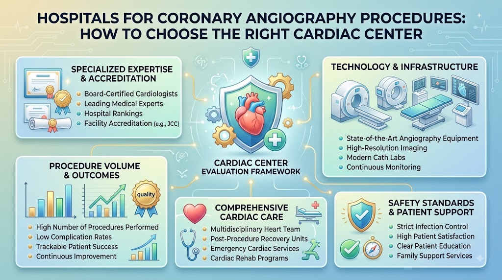
When evaluating hospitals for coronary angiography procedures, look for centers that maintain high standards across clinical staff, facilities, and emergency protocols.
                  ┌──────────────────────────────────────────┐
                  │    Core Elements of a Cardiac Center     │
                  └─────────────────────┬────────────────────┘
                                        │
      ┌──────────────────┬──────────────┴───────────────┬──────────────────┐
      ▼                  ▼                              ▼                  ▼
┌───────────┐    ┌──────────────┐              ┌───────────────┐    ┌──────────────┐
│ Clinical  │    │  Cath Lab    │              │ Advanced      │    │ Emergency &  │
│ Team      │    │  Facilities  │              │ Imaging       │    │ Surgical Care│
└───────────┘    └──────────────┘              └───────────────┘    └──────────────┘
Experienced Multidisciplinary Cardiology Team
A top-tier cardiac program relies on a collaborative team of specialists:
- Interventional Cardiologists: Subspecialized cardiologists with advanced training in catheter-based diagnostic and therapeutic procedures.
- Clinical Cardiologists: Physicians who manage overall cardiovascular health, interpret initial diagnostics, and direct post-procedure care.
- Cardiac Anesthesiologists: Specialists available for patients with complex medical conditions or severe anxieties who require monitoring during catheterization.
- Cardiothoracic Surgeons: On-site surgical support if diagnostic findings require surgical evaluation or emergency intervention.
- Cath Lab Nurses and Radiologic Technologists: Staff specifically trained in sterile catheterization techniques, hemodynamics, radiation safety, and acute cardiac care.
Modern Cardiac Catheterization Laboratory (Cath Lab)
The Cath Lab is the specialized procedure suite where your angiogram takes place. Look for hospitals that feature dedicated, climate-controlled catheterization rooms equipped with low-dose radiation fluoroscopy systems, continuous hemodynamic monitoring tools, and integrated intravascular imaging options (such as IVUS or OCT).
Advanced Diagnostic & Imaging Capabilities
High-quality facilities offer on-site access to complementary diagnostic infrastructure:
- Dual-source high-resolution CT scanners.
- Transthoracic and Transesophageal Echocardiography (TTE/TEE).
- Point-of-care blood testing laboratories for rapid monitoring of cardiac enzymes, clotting times, and kidney function markers.
Emergency Cardiac Support & Intensive Care
Because diagnostic catheterization is an invasive procedure, the hospital must be prepared for rare emergency situations:
- A dedicated Intensive Cardiac Care Unit (ICCU) staffed 24/7 by critical care cardiologists.
- Immediate access to advanced circulatory support, such as intra-aortic balloon pumps (IABP) or Impella devices.
- Rapid-response protocols for transition to emergency angioplasty or surgical suites.
What Is a Cardiac Cath Lab?
A Cardiac Catheterization Laboratory (Cath Lab) is a specialized, sterile procedure room designed for minimally invasive heart procedures. Unlike standard surgical operating rooms, a Cath Lab is built around dynamic X-ray imaging systems (C-arms) that move around the patient table to capture high-definition, multi-angle images of the beating heart.
                           [ C-Arm X-Ray System ]
                                     │
                                     ▼
[ Sterile Patient Table ] ───> [ Catheter Insertion ] ───> [ Live High-Def Monitors ]
                                     │                      (Physician tracks dye
                                     ▼                       flow in real time)
                       [ Hemodynamic Control Unit ]
                       (Continuous vital sign monitoring)
In the Cath Lab:
- Physicians guide delicate catheters through blood vessels using real-time fluoroscopic displays.
- Hemodynamic monitoring systems continuously display heart rate, arterial blood pressure, blood oxygenation, and intracardiac pressure waves.
- Sterile conditions prevent infection risks at the arterial insertion site.
While the primary role of a Cath Lab during an angiogram is diagnostic, the lab is equipped so that if an actionable, life-threatening blockage is identified, the team can transition—with patient consent—to therapeutic interventional procedures like angioplasty or stent placement.
The Step-by-Step Coronary Angiography Procedure
Knowing what happens during a coronary angiogram can help alleviate anxiety. The procedure generally follows a standard clinical progression:
┌────────────────────────────────────────────────────────────────────────┐
│                   The Angiography Patient Journey                      │
├────────────────────────────────────────────────────────────────────────┤
│ Step 1: Pre-Procedure Clinical & Laboratory Assessment                 │
│ Step 2: Admission, Prep, & Intravenous (IV) Line Placement             │
│ Step 3: Local Anesthetic Injection at Access Site (Wrist/Groin)        │
│ Step 4: Sheath & Catheter Insertion Under Fluoroscopic Guidance        │
│ Step 5: Contrast Dye Injection & Dynamic X-Ray Imaging                 │
│ Step 6: Catheter Removal & Hemostasis (Pressure Application)          │
│ Step 7: Post-Procedure Recovery, Observation, & Results Review         │
└────────────────────────────────────────────────────────────────────────┘
Step 1: Pre-Procedure Assessment
Before entering the Cath Lab, the clinical staff reviews your medical history, verifies your identity, confirms fasting status, checks blood tests (specifically kidney markers like serum creatinine and blood clotting profiles), and secures formal informed consent.
Step 2: Preparing the Patient
You will change into a hospital gown, and an IV line will be placed in your arm to administer fluids or mild sedatives if needed. Nurses will shave and disinfect the chosen access site (wrist or groin) and connect ECG patches to track your heart rhythm continuous.
Step 3: Local Anesthesia
You are positioned comfortably on the padded Cath Lab table. The clinical team cleans the entry area with antiseptic solution and drapes you in sterile sheets. The cardiologist injects a local anesthetic into the skin over your radial (wrist) or femoral (groin) artery. You may feel a brief pinch or sting before the area goes numb.
Step 4: Catheter Insertion
A small puncture is made in the numbed artery, and a short guide sheath is positioned. The physician inserts a soft-tipped catheter into the sheath, advancing it through your vascular system up to the coronary arteries. Because the insides of blood vessels lack nerve endings for pain, you will not feel the catheter moving through your body.
Step 5: Contrast Injection & Imaging
Once the catheter tip is located at the entrance of a coronary artery, the physician injects a small volume of radio-opaque contrast dye. You might feel a momentary warm or flushing sensation spread through your chest or body—this is a normal reaction to the contrast medium. Dynamic X-ray equipment captures video images as the dye fills the arterial branches.
Step 6: Catheter Removal & Hemostasis
After completing the imaging, the physician withdraws the catheter and sheath. The entry site requires immediate pressure to prevent bleeding:
- Radial Access (Wrist): A specialized compression wristband is applied to exert gentle, controlled pressure over the puncture site.
- Femoral Access (Groin): Manual pressure is applied for 15–30 minutes, or a specialized vascular closure device (such as a plug or suture) is used to seal the artery.
Step 7: Results and Next Steps
The diagnostic images are reviewed immediately by the interventional cardiologist. Findings are shared with you and your family to decide the next steps, whether that means continued medical management, schedule adjustments, or further interventional procedures.
Procedure Duration and Hospital Stay Expectations
Understanding the timeline helps set realistic expectations for your procedure day:
- Active Procedure Time: The actual angiography inside the Cath Lab typically takes between 20 to 45 minutes.
- Total Preparation & Recovery Time: Factoring in pre-procedure prep, sterile draping, site compression, and post-procedure recovery, the entire process takes 4 to 8 hours.
+--------------------------------------------------------------------+
|                      SAME-DAY DISCHARGE CONDITIONS                 |
+--------------------------------------------------------------------+
|  Many uncomplicated diagnostic angiograms—especially those         |
|  performed via the radial (wrist) approach—are completed as day-   |
|  case procedures, allowing patients to return home the same day.   |
|                                                                    |
|  Overnight hospital admission may be advised if:                   |
|   • Access was gained through the groin (requiring longer bed rest)|
|   • You have significant co-existing medical conditions            |
|   • An immediate angioplasty or stent placement was performed      |
|   • You experience post-procedure discomfort or delayed bleeding   |
+--------------------------------------------------------------------+
Preparing for Your Coronary Angiography
Proper preparation minimizes procedural risks and helps ensure clear imaging results. Follow all specific prep guidelines provided by your hospital care team.
┌─────────────────────────────────────────────────────────────────┐
│                    Patient Preparation Steps                    │
├───────────────────┬─────────────────────────┬───────────────────┤
│ Fasting Guidelines│ Medication Review       │ Logistical Prep   │
├───────────────────┼─────────────────────────┼───────────────────┤
│ Fast from solid   │ Review all blood        │ Arrange for a     │
│ foods for 4 to 8  │ thinners, diabetes      │ responsible adult │
│ hours before the  │ medications, and ACE    │ driver to take    │
│ procedure         │ inhibitors with doctors │ you home          │
└───────────────────┴─────────────────────────┴───────────────────┘
Crucial Medication Safety Warning: Never stop, start, or alter your prescribed medication doses on your own. Adjustments to blood thinners (e.g., Warfarin, Clopidogrel, NOACs) or diabetes medications (especially Metformin) must only be made under the direct instruction of your treating cardiology team.
Pre-Procedure Disclosure Checklist
Before entering the Cath Lab, ensure your clinical team is fully informed of your medical background. Use this checklist during your pre-procedure consultation:
 [ ] History of allergic reactions to iodine-based contrast dyes or shellfish
 [ ] Known kidney disease, elevated creatinine levels, or single-kidney status
 [ ] Complete list of prescription drugs, OTC supplements, and herbal remedies
 [ ] Current intake of antiplatelet or anticoagulant (blood-thinning) medications
 [ ] Diagnosis of diabetes and current insulin/Metformin regimens
 [ ] History of bleeding disorders or unexplained bruising issues
 [ ] Previous vascular, bypass, or cardiac valve operations
 [ ] Possibility of pregnancy or active breastfeeding (for female patients)
 [ ] Recent fevers, infections, or acute physical illness
Understanding the Risks of Coronary Angiography
Coronary angiography is a routinely performed, safe procedure. However, as an invasive diagnostic test involving catheters and blood vessels, it carries potential risks. NHS guidelines and cardiology standards summarize potential complications as follows:
┌─────────────────────────────────────────────────────────────────────────┐
│                    Potential Angiography Complications                  │
├───────────────────────────────────┬─────────────────────────────────────┤
│ Minor / Relatively Common Effects  │ Rare to Very Severe Complications   │
├───────────────────────────────────┼─────────────────────────────────────┤
│ • Minor bruising or discoloration │ • Severe bleeding at puncture site  │
│   at the arterial entry site      │ • Blood vessel damage or tear       │
│ • Localized soreness or tenderness│ • Contrast-induced kidney stress    │
│ • Small, self-limiting hematoma   │ • Anaphylactic contrast allergy     │
│ • Temporary flushing from contrast│ • Heart attack, stroke, or arrhythmia│
└───────────────────────────────────┴─────────────────────────────────────┘
Complication rates vary depending on age, kidney health, emergency status, and underlying heart function. Discuss your individual risk profile with your cardiologist before the procedure.
Coronary Angiography and Kidney Health
The contrast dye used during angiography is filtered and eliminated from your body by your kidneys. In some individuals, iodine-based contrast media can temporarily strain renal function—a condition known as Contrast-Induced Nephropathy (CIN) or Contrast-Induced Acute Kidney Injury (CI-AKI).
                 [ Contrast Agent Injected ]
                              │
                              ▼
                 [ Eliminated via Kidneys ]
                              │
            ┌─────────────────┴─────────────────┐
            ▼                                   ▼
[ Normal Kidney Function ]          [ Impaired Kidney Function ]
Risk is very low; body               Requires clinical precautions:
flushes dye naturally                • Pre-procedure hydration
                                     • Minimal contrast volume
                                     • Post-procedure blood monitoring
- Risk Evaluation: Your medical team will order a baseline blood test (Serum Creatinine and Estimated Glomerular Filtration Rate – eGFR) before scheduling the test.
- Preventive Measures: If you have reduced kidney function, the team may administer IV fluids before and after the test to help your kidneys flush the dye. They will also select low-osmolar contrast agents and minimize dye volume.
Always inform your care team if you have a history of kidney disease or are taking medications that affect kidney function.
What Happens If a Blockage Is Identified?
An angiogram provides a clear visual map of your coronary anatomy. The findings guide your cardiologist in recommending the next steps for your care.
                         [ Angiogram Completed ]
                                    │
         ┌──────────────────────────┼──────────────────────────┐
         ▼                          ▼                          ▼
[ Clear / Minimal CAD ]     [ Moderate Narrowing ]    [ Severe / Multi-Vessel ]
  No major blockages         Single or localized        Complex or multi-artery
  found.                     narrowing.                 blockages.
         │                          │                          │
         ▼                          ▼                          ▼
Medical Management          Possible Angioplasty       Surgical Consultation
(Lifestyle & Meds)          & Stent Placement          (CABG / Bypass Surgery)
- Clear or Minimal Disease: If the arteries show no significant narrowing, treatment typically focuses on managing risk factors like cholesterol, blood pressure, and lifestyle habits.
- Moderate Narrowing: If one or two localized blockages are found, your physician may recommend opening the vessel with a balloon and placing a wire mesh tube called a stent (angioplasty). In many cases, if consent was provided ahead of time, this can be performed during the same Cath Lab session.
- Severe or Multi-Vessel Disease: If multiple arteries are severely narrowed or the Left Main coronary artery is involved, your team may recommend a Coronary Artery Bypass Graft (CABG) surgery. The findings will be reviewed by a Heart Team (including interventional cardiologists and cardiac surgeons) to determine the safest approach.
Clinical Reality: Identifying an arterial blockage during an angiogram does not automatically mean you need a stent or bypass surgery. The best treatment depends on your symptoms, the location and severity of the blockage, and your overall heart health.
Angiography vs. Angioplasty: Key Differences
It is common to confuse these two terms. While both are performed in a Cath Lab using arterial catheters, their clinical objectives are distinct:
┌────────────────────────────────────────────────────────────────────────┐
│                   Angiography vs. Angioplasty                          │
├───────────────────────────────┬────────────────────────────────────────┤
│ Feature                       │ Coronary Angiography                   │
├───────────────────────────────┼────────────────────────────────────────┤
│ Primary Purpose               │ Diagnostic (visualizing blood vessels) │
│ Interventional Tools Used     │ Catheter and contrast dye              │
│ Procedure Result              │ Detailed image map of arteries         │
├───────────────────────────────┼────────────────────────────────────────┤
│ Feature                       │ Coronary Angioplasty (PCI)             │
├───────────────────────────────┼────────────────────────────────────────┤
│ Primary Purpose               │ Therapeutic (opening blocked vessels)  │
│ Interventional Tools Used     │ Balloons, stents, atherectomy devices  │
│ Procedure Result              │ Restored blood flow through artery     │
└───────────────────────────────┴────────────────────────────────────────┘
An angiography gathers information to make a diagnosis. An angioplasty is the intervention used to treat a discovered blockage.
How to Choose the Right Cardiac Hospital
Selecting an appropriate hospital for a coronary angiography procedure involves evaluating key quality, safety, and operational standards. Use this criteria list to guide your decision:
┌────────────────────────────────────────────────────────────────────────┐
│                      Hospital Evaluation Criteria                      │
├────────────────────────────────────────────────────────────────────────┤
│  1. Accredited Cardiac Facilities (NABH, JCI, or national equivalents) │
│  2. Board-certified Interventional Cardiologists on staff              │
│  3. Modern, high-definition Cath Lab equipment                         │
│  4. 24/7 Intensive Cardiac Care Unit (ICCU) support                    │
│  5. On-site Cardiothoracic Surgery capabilities                        │
│  6. Protocols for pre-procedure kidney risk screening                  │
│  7. Low rate of procedural complications and site infections           │
│  8. Transparent pricing with clear estimates                           │
│  9. Insurance network partnership and billing support                   │
│ 10. Clear discharge instructions and formal follow-up plans            │
└────────────────────────────────────────────────────────────────────────┘
Cost Components of Coronary Angiography
The total cost of a coronary angiography procedure can vary based on several factors, including location, hospital facility type, and individual patient needs.
┌─────────────────────────────────────────────────────────────────────────┐
│                     Angiography Cost Breakdown                          │
├───────────────────────┬─────────────────────────────────────────────────┤
│ Fee Component         │ What Is Included                                │
├───────────────────────┼─────────────────────────────────────────────────┤
│ Procedure Fee         │ Core charge for Cath Lab room and equipment     │
│ Specialist Fee        │ Professional fee for the interventional doctor  │
│ Consumables & Contrast│ Sterile catheters, sheaths, and contrast dye    │
│ Diagnostic Pre-Tests  │ Blood tests (creatinine, clotting), ECG, echo   │
│ Day-Care / Room Rent  │ Recovery room, observation bed, nursing care    │
│ Medication Charges    │ IV fluids, local anesthesia, discharge medicines│
└───────────────────────┴─────────────────────────────────────────────────┘
Key Factors Influencing Cost Differences
- Access Route & Technology: Radial access may reduce recovery time and room charges, while specialized closure devices or complex imaging can add to the baseline fee.
- Admit Status vs. Day Care: A day-case procedure is generally less expensive than an overnight hospital stay.
- Additional Interventions: If an unplanned angioplasty or stent placement is required during the procedure, overall costs will increase significantly due to specialized equipment and longer observation times.
Cost Advice: Always request a detailed, written cost breakdown from the hospital billing department prior to the procedure. Be sure to clarify what is included in the base package and what might trigger additional charges.
Insurance and Financial Planning Considerations
Navigating insurance coverage for diagnostic cardiac procedures helps prevent unexpected out-of-pocket expenses:
- Pre-Authorization: Most health insurance plans require pre-authorization for elective invasive procedures. Confirm that your hospital’s administrative team submits all necessary clinical documentation well ahead of time.
- In-Network Benefits: Check if the cardiac hospital and attending cardiologist are within your insurer’s cashless network to simplify billing.
- Consumables and Imaging: Confirm whether contrast dye, catheters, and post-procedure recovery room stays are fully covered under your policy’s diagnostic benefit clauses.
- Contingency Approval: Ask your insurer how coverage works if a diagnostic angiogram transitions directly into an emergency or planned angioplasty during the same session.
What to Expect After the Procedure
Recovery after an angiogram depends primarily on the arterial access site used during your test:
┌─────────────────────────────────────────────────────────────────────────┐
│                     Post-Procedure Recovery Care                        │
├───────────────────────────────────┬─────────────────────────────────────┤
│ Radial Access (Wrist)             │ Femoral Access (Groin)              │
├───────────────────────────────────┼─────────────────────────────────────┤
│ • Compression band applied for    │ • Firm manual pressure or closure   │
│   2 to 4 hours                    │   device applied to access site     │
│ • Sitting up and walking is       │ • Requires strict flat bed rest for │
│   usually permitted early         │   4 to 6 hours to prevent bleeding  │
│ • Keep wrist rested and dry       │ • Avoid bending at the waist or     │
│   for the first 24–48 hours       │   straining during initial recovery │
└───────────────────────────────────┴─────────────────────────────────────┘
General Discharge Guidelines
- Hydration: Drink plenty of fluids (as advised by your care team) over the next 24 hours to help flush the contrast dye from your system.
- Activity Limits: Avoid heavy lifting, strenuous exercise, driving, or vigorous exertion for a few days post-procedure.
- Site Care: Keep the puncture site clean and dry. Inspect it daily for signs of swelling, redness, or bruising.
Post-Procedure Warning Signs
While serious complications are uncommon, you should contact your hospital emergency line or seek immediate medical care if you experience any of the following warning signs:
[ CRITICAL WARNING SIGNS - SEEK IMMEDIATE MEDICAL ATTENTION ]
 • Active, bright red bleeding or rapid swelling at the puncture site
 • Severe, worsening pain, coldness, numbness, or tingling in the arm or leg used
 • Sudden chest pain, pressure, or shortness of breath
 • Fever, chills, or spreading redness/warmth around the insertion site
 • Feeling faint, dizzy, unusually lightheaded, or confused
 • Significant decrease in urine output or unusually dark urine
How to Compare Cardiac Hospitals
Using a structured comparison table can help you evaluate different hospitals objectively based on verified clinical information:
| Comparison Factor | Hospital A | Hospital B | Notes / Verification |
| Cath Lab Availability | 24/7 Dedicated Suite | Standard Shared Room | Look for dedicated cardiac suites |
| Primary Access Route | Radial (Wrist) Preferred | Femoral (Groin) Standard | Radial often allows faster recovery |
| Cardiology Experience | Board-Certified Interventional | General Cardiology | Verify interventional credentials |
| On-Site Cardiac Surgery | Full Surgical Team | On-Call / Transfer | Important for complex disease |
| Intensive Care Unit | Dedicated ICCU | General ICU | Dedicated ICCU offers focused care |
| Renal Safety Protocols | Pre-Test eGFR Hydration | Standard Protocol | Crucial for patients with kidney disease |
| Billing Transparency | Itemized Written Estimate | Lump-Sum Estimate | Clarify inclusions for contrast/equipment |
| Insurance Coverage | In-Network (Cashless) | Out-of-Network | Confirm pre-authorization rules |
Hospital Selection Red Flags
Be cautious of health facilities that exhibit any of these procedural or operational warning signs:
┌─────────────────────────────────────────────────────────────────────────┐
│                           WARNING RED FLAGS                             │
├─────────────────────────────────────────────────────────────────────────┤
│ ✖ Promising "100% safe" or completely risk-free invasive procedures     │
│ ✖ Guaranteeing specific blockage findings before imaging is done        │
│ ✖ Pressuring you into immediate elective stenting without clear rationale│
│ ✖ Refusing to provide written cost estimates or itemized billing details│
│ ✖ Lacking on-site emergency facilities or critical care support        │
│ ✖ Unwillingness to discuss doctor qualifications or Cath Lab standards  │
│ ✖ Omitting pre-procedure kidney tests or health history reviews         │
└─────────────────────────────────────────────────────────────────────────┘
Common Myths vs. Medical Facts
┌─────────────────────────────────────────────────────────────────────────┐
│                           MYTHS VS. FACTS                               │
├───────────────────────────────────────────┬─────────────────────────────┤
│ MYTH                                      │ FACT                        │
├───────────────────────────────────────────┼─────────────────────────────┤
│ "An angiogram automatically means you     │ "Angiography is a diagnostic│
│  will get a stent."                       │  test; treatment depends on │
│                                           │  the visual results."       │
├───────────────────────────────────────────┼─────────────────────────────┤
│ "Coronary angiography is major open-heart │ "It is a minimally invasive │
│  surgery."                                │  catheter procedure using   │
│                                           │  local anesthesia."         │
├───────────────────────────────────────────┼─────────────────────────────┤
│ "Every patient needs the exact same       │ "Preparation varies based on│
│  preparation and recovery schedule."      │  medical history, access    │
│                                           │  site, and kidney function."│
├───────────────────────────────────────────┼─────────────────────────────┤
│ "The cheapest hospital is always the best │ "Price should be evaluated  │
│  choice for diagnostic procedures."       │  alongside Cath Lab quality │
│                                           │  and emergency support."    │
├───────────────────────────────────────────┼─────────────────────────────┤
│ "You must go under general anesthesia     │ "Most procedures use local  │
│  for an angiogram."                       │  anesthetic, allowing you to│
│                                           │  stay awake and comfortable."│
└───────────────────────────────────────────┴─────────────────────────────┘
Common Patient Mistakes to Avoid
- Choosing Based Solely on Cost: Selecting a facility based only on low prices without reviewing Cath Lab standards, specialist experience, or emergency capabilities.
- Hiding Medical History: Omitting details about contrast dye allergies, kidney conditions, or over-the-counter medications during pre-procedure evaluation.
- Adjusting Medications Independently: Discontinuing blood thinners or diabetes medications without explicit guidance from your cardiology team.
- Assuming Stenting Is Mandatory: Believing that any identified arterial narrowing automatically requires immediate stent placement or open-heart surgery.
- Ignoring Post-Discharge Instructions: Resuming heavy physical exertion too soon or neglecting puncture site care guidelines.
How BESTCARDIACHOSPITALS Can Support Your Decision
Finding clear, dependable information is an important part of managing your cardiovascular care. BESTCARDIACHOSPITALS.COM offers patient-focused resources to help you evaluate cardiac facilities and prepare for your appointments:
- Hospital Evaluation Criteria: Learn how to assess Cath Lab infrastructure, specialist credentials, and emergency support.
- Procedure Educational Guides: Understand common cardiac tests, interventional treatments, and recovery protocols.
- Discussion Checklists: Access structured questions to help you have informed conversations with your care team.
- Comparative Insights: Learn about essential safety standards, billing practices, and accreditation benchmarks.
Frequently Asked Questions (FAQs)
1.What is a coronary angiography procedure?
A coronary angiography is an invasive diagnostic test that uses X-ray fluoroscopy and contrast dye to visualize the inside of your coronary arteries and check for blockages or narrowing.
2.Why is coronary angiography performed?
It is performed to investigate unexplained chest pain, evaluate abnormal results from non-invasive tests (like stress tests), assess coronary artery disease, and plan potential treatments like angioplasty or surgery.
3.How do I choose the right hospital for coronary angiography?
Look for facilities with accredited Cath Labs, experienced interventional cardiologists, on-site ICCU emergency backup, cardiothoracic surgical capabilities, transparent pricing, and clear kidney safety protocols.
4.What is a cardiac catheterization laboratory (Cath Lab)?
A Cath Lab is a specialized, sterile procedure room equipped with high-resolution C-arm X-ray systems and continuous vital sign monitoring designed for catheter-based diagnostic and therapeutic heart procedures.
5.How long does a coronary angiogram take?
The active diagnostic imaging inside the Cath Lab generally takes 20 to 45 minutes. Total hospital preparation and recovery time usually ranges from 4 to 8 hours.
6.Is coronary angiography painful?
Local anesthetic numbs the catheter insertion site (wrist or groin) to ensure comfort. You remain awake and may feel slight pressure during catheter placement or a brief warm sensation when contrast dye is injected.
7.How should I prepare for my coronary angiogram?
Follow your care team’s fasting instructions (usually no solid food for 4 to 8 hours prior), provide a complete list of your medications and allergies, and arrange for someone to drive you home.
8.Can I take my regular medications before angiography?
Certain medications (such as blood thinners or diabetes drugs like Metformin) may need to be temporarily paused or adjusted. Never change your dosage without explicit instructions from your treating physician.
9.What are the main risks associated with coronary angiography?
Common minor risks include local bruising or tenderness at the insertion site. Rare, serious risks include bleeding, blood vessel damage, contrast-induced kidney stress, allergic reactions, or cardiovascular complications.
10.Can coronary angiography detect all arterial blockages?
Yes, angiography provides direct, real-time visual mapping of the internal channels of your coronary arteries, allowing physicians to pinpoint the location and severity of blockages.
11.Does an angiogram always require stent placement?
No. An angiogram is primarily a diagnostic test. Findings may lead to recommendations for medical management, lifestyle changes, angioplasty with stenting, or bypass surgery, depending on the severity and location of any blockages.
12.How much does coronary angiography cost?
Costs vary based on the facility, doctor fees, geographic location, choice of access site, contrast dye usage, and whether the procedure is performed as a day case or requires an overnight stay. Always request a detailed written estimate from the hospital.
13.What is the typical recovery time after an angiogram?
Many patients undergoing radial (wrist) access resume light activities within 24 to 48 hours. Femoral (groin) access may require a slightly longer rest period to ensure the entry site heals properly.
14.What should I ask my cardiologist before the procedure?
Ask about their experience, whether they plan to use radial access, pre-procedure kidney checks, overall risks based on your health history, and what steps will be taken if blockages are found.
15.What warning signs require immediate medical attention after discharge?
Seek emergency care for active bleeding or rapid swelling at the insertion site, severe arm or leg pain, coldness in the limb, sudden chest pain, fever, or shortness of breath.
Final Conclusion
A coronary angiography is a helpful diagnostic procedure that offers dynamic, real-time images of your heart’s blood supply. While receiving a recommendation for an angiogram can feel overwhelming, understanding how the procedure works empowers you to take an active role in your care.