Your heart is at the center of your health, and when it’s not functioning optimally, it can impact every part of your life. Angioplasty, a groundbreaking procedure, is one of the most effective ways to restore blood flow to your heart, ensuring it continues to beat strong and steady. At Best Cardiac Hospitals, we specialize in performing angioplasty with precision, care, and cutting-edge technology, all tailored to your unique needs.
Whether you're seeking to understand the procedure or preparing for it, this page will provide you with all the information you need to feel confident and informed.
What is Angioplasty?
Angioplasty, also known as Percutaneous Coronary Intervention (PCI),
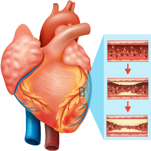
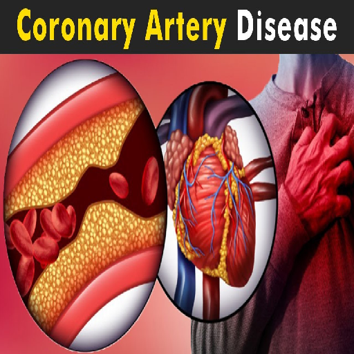
is a medical procedure designed to treat blocked or narrowed coronary arteries. These arteries are vital for delivering oxygen-rich blood to the heart. When they become obstructed, it can result in symptoms such as chest pain (angina), shortness of breath, or even severe conditions like a heart attack. As a minimally invasive technique, angioplasty is widely used to restore blood flow and alleviate these potentially life-threatening complications.
The procedure involves inserting a thin, flexible tube called a catheter into a blood vessel and guiding it
to the blocked artery. Once in place, a small balloon at the tip of the catheter is inflated to widen the artery. In most cases, a stent, a tiny mesh tube, is inserted to maintain the artery's openness and ensure blood can flow freely. This combination of balloon inflation and stent placement has proven effective in improving heart health and preventing future cardiac events.
Types of Angioplasty
Every heart and case of coronary artery disease is unique, requiring personalized approaches
to treatment. Angioplasty offers several techniques to address varying needs and ensure effective outcomes. Below are the primary types of angioplasty and their specific purposes:
Balloon Angioplasty
This is one of the simplest forms of angioplasty. A small balloon at the tip of a catheter is guided to
the blockage in the coronary artery. Once in place, the balloon is inflated to compress the plaque against the artery walls, widening the passage for blood flow. Although highly effective, this method often requires additional measures, such as stent placement, to ensure long-term results.
Stent Placement
Stent placement is a commonly performed procedure in conjunction with angioplasty. Once the artery is
widened using the balloon, a stent—a small wire-mesh tube—is inserted to keep it open. Stents offer crucial support, preventing the artery from collapsing or narrowing again. Two types of stents are commonly used:
Drug-Eluting Stents (DES): These stents are coated with medication that is slowly
released over time to minimize the risk of restenosis (re-narrowing of the artery).
Bare-Metal Stents (BMS): These are uncoated stents that provide structural support
to the artery. While effective, they may carry a slightly higher risk of restenosis compared to drug-eluting stents.
Laser Angioplasty
For particularly stubborn or complex blockages, laser angioplasty provides an advanced alternative.
This technique involves the use of laser energy to vaporize the plaque buildup in the artery. It’s a highly precise method, making it suitable for hard-to-reach or densely calcified areas. Laser angioplasty is often used in combination with other techniques to achieve optimal results.
Rotational Atherectomy
In some cases, when the plaque is calcified and too hard for standard methods, a rotational atherectomy
may be performed. This involves using a tiny, diamond-tipped drill to grind away the hardened plaque. Once the artery is sufficiently cleared, other techniques like stent placement can follow.
Cutting Balloon Angioplasty
This specialized form of angioplasty employs a balloon with small blades or scoring edges. As the
balloon inflates, the blades make precise incisions in the plaque, allowing it to be compressed more effectively. This method is particularly useful for treating blockages that are resistant to standard balloon angioplasty.
How is Angioplasty Performed?
The angioplasty procedure is typically done in a specialized area called a catheterization lab (cath lab). Here’s what you can expect:
1. Preparing for the Procedure
Local Anesthesia: During the procedure, local anesthesia ensures you remain awake
while eliminating any sensation of pain at the insertion site. This method allows for patient comfort and avoids the risks associated with general anesthesia. You’ll be able to communicate with the medical team throughout the process.
Catheter Insertion: A small incision is made in the groin, wrist, or arm to access an
artery, providing a pathway for the catheter. This thin, flexible tube is skillfully inserted and guided through the blood vessels using imaging technology. The catheter's precision placement allows for safe navigation to the heart for treatment.
2. Balloon Inflation
Reaching the Blockage: Advanced imaging techniques, such as X-rays or fluoroscopy, are used
to guide the catheter through the blood vessels to the exact location of the blockage. This ensures the procedure is highly precise and minimizes risks to surrounding areas. The catheter's position is carefully monitored to ensure it reaches the affected artery safely.
Widening the Artery: Once the catheter is in place, a small balloon at its tip is inflated
at the site of the blockage. The inflated balloon compresses the plaque buildup against the artery wall, creating a wider pathway for blood flow. This critical step restores normal circulation, reducing symptoms and preventing further heart complications.
3. Stent Placement (if needed)
In most cases, a stent, mesh-like tube—is placed at the site of the blockage to ensure the artery remains open. This stent acts as a scaffold, preventing the artery from narrowing again over time. Stent placement is a crucial step in maintaining long-term blood flow and reducing the likelihood of requiring repeat procedures.
4. Final Steps
Ensuring Success: Once the artery is confirmed to be successfully opened and blood flow
is restored, the catheter is carefully withdrawn. The medical team ensures the procedure has achieved its goal, and the incision site is securely closed to prevent bleeding or infection. This marks the completion of the intervention with minimal disruption to the body.
Hospital Stay: Following the procedure, most patients stay in the hospital for
observation overnight. This allows healthcare professionals to monitor vital signs, ensure there are no complications, and provide necessary post-operative care. The observation period helps ensure a smooth recovery before the patient is discharged.
What Happens During Angioplasty?
Undergoing a medical procedure like angioplasty can be intimidating, but understanding each step can provide reassurance and help you feel more prepared. The process is typically straightforward and involves three key phases: preparation, the procedure itself, and recovery.
Before the Procedure
Before angioplasty, your doctor will perform a thorough evaluation of your heart health. This includes diagnostic tests such as blood work, electrocardiograms (EKG), and imaging studies like coronary angiography to pinpoint the location and severity of the blockage. You may be asked to fast for a few hours before the procedure to ensure the process goes smoothly. If you’re on specific medications, your doctor may adjust the dosages or provide special instructions. Additionally, you’ll have a chance to discuss any concerns or questions with your medical team, ensuring you’re well-informed and at ease.
During the Procedure
Angioplasty is typically performed in a catheterization lab, a specialized operating room equipped for such procedures. You’ll remain awake but sedated to minimize discomfort while maintaining awareness. A small incision is made in either your wrist or groin to access a blood vessel. A thin, flexible tube called a catheter is carefully guided through the blood vessels using X-ray imaging until it reaches the blocked artery. Once positioned, a tiny balloon at the catheter's tip is inflated to compress the plaque and widen the artery. In most cases, a stent—a small wire mesh tube—is inserted to keep the artery open and maintain proper blood flow. The procedure usually lasts about one to two hours, depending on the complexity of the blockage.
After the Procedure
Following angioplasty, recovery begins almost immediately. You’ll be monitored in the hospital for a few hours or overnight to ensure there are no complications. The access site, whether in the wrist or groin, will be checked for bleeding or swelling. Your doctor will provide post-procedure care instructions, including medications to prevent blood clots and lifestyle recommendations to improve heart health. Most patients can resume normal activities within a few days, but heavy lifting and strenuous exercise may be restricted temporarily. A follow-up appointment will be scheduled to monitor your recovery and the success of the angioplasty.
What to Expect After Angioplasty
After angioplasty, most patients recover quickly, typically leaving the hospital within
24 hours and resuming light activities soon after. Regular follow-ups ensure the stent is functioning properly, while lifestyle changes like a heart-healthy diet, regular exercise, and stress management are key to long-term success. Quitting smoking and adopting these habits significantly reduce the risk of future blockages and improve overall heart health.
Recovery Process
Hospital Stay: Patients are usually discharged within 24 hours after the procedure.
Activity Restrictions: Avoid strenuous activities for a few days, but walking and light
movements are encouraged.
Follow-Up Care: Regular check-ups ensure the stent is functioning well and there are
no complications.
Lifestyle Adjustments
Adopt a heart-healthy diet rich in fruits, vegetables, whole grains, and lean proteins.
Engage in regular exercise, as recommended by your doctor.
Quit smoking and manage stress to reduce your risk of future blockages.
Risks and How They’re Managed
Like any procedure, angioplasty has risks, but modern techniques and experienced
cardiologists make complications rare. Potential risks include:
Bleeding or Bruising
At the catheter insertion site, swelling, or tenderness.
Artery Re-Narrowing
Particularly with bare-metal stents.
Blood Clots
Managed with prescribed blood thinners.
Allergic Reactions
Rare, but possible with the dye used for imaging.
Frequently Asked Questions (FAQ)
Q: Is angioplasty painful?
A: Not at all. You’ll be under local anesthesia and may feel some pressure but no pain.
Q: How long does recovery take?
A: Most people resume daily activities within a week, though full recovery may take a few weeks.
Q: Can angioplasty cure heart disease?
A: While angioplasty treats blockages, maintaining a healthy heart requires lifestyle changes and regular check-ups.
Q: Will I need angioplasty again?
A: With proper care and medication, repeat procedures are often unnecessary.
Q: Is angioplasty safe for older adults?
A: Yes, angioplasty is commonly performed on seniors with excellent outcomes.
Ready to Take the Next Step?
Don’t let fear hold you back from living your best life. Whether you’re facing symptoms or seeking preventive care, angioplasty can pave the way to a healthier future. Connect with the world’s leading cardiac hospitals through our platform and take control of your heart health today.
Top Hospitals Top Surgery Top Destinations Top Surgeons